Category Archives: Abdomen

Liver Laceration And Liver Function Tests

Over the years I’ve seen a number of trauma professionals, both surgeons and emergency physicians, order liver transaminases (SGOT, SGPT) and bilirubin in patients with liver laceration. I’ve never been clear on why, so I decided to check it out. As it turns out, this is another one of those “old habits die hard” phenomena.
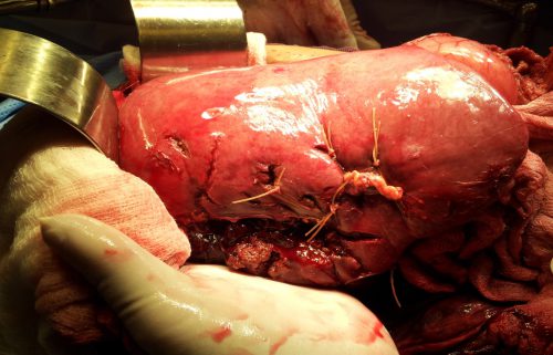
Liver lacerations, by definition, are disruptions of the liver parenchyma. Liver tissue and bile ducts of various size are both injured. Is it reasonable to expect that liver function tests would be elevated? A review of the literature follows the typical pattern. Old studies with very few patients.

From personal hands-on observations, the liver tissue itself tears easily, but the ducts are a lot tougher. It is fairly common to see small, intact ducts bridging small tears in the substance of the liver. However, larger injuries can certainly disrupt major ducts, leading to major problems. But I’ve never seen obstructive problems develop from this injury.

A number of papers (very small, retrospective series) have shown that transaminases can rise with liver laceration. However, they do not rise reliably enough to be a good predictor of either having an injury, or the degree of injury. Similarly, bilirubin can be elevated, but usually not as a direct result of the injury. The most common causes are breakdown of transfused or extravasated blood, or from critical care issues like sepsis, infection, and shock.

Bottom line: Don’t bother to get liver function tests in patients with known or suspected injury. Only a CT scan can help you find and/or grade the injury. And never blame an elevated bilirubin on the injury. Start searching for other causes, because they will end up being much more clinically significant.

References:

  • Evaluation of liver function tests in screening for intra-abdominal injuries. Ann Emerg Med 20(8):838-841, 1991.
  • Markers for occult liver injury in cases of physical abuse in children. Pediatrics 89(2):274-278.
  • Combination of white blood cell count with liver enzymes in the diagnosis of blunt liver laceration. Am J Emerg Med 28(9):1024-1029, 2010.

Blunt Duodenal Injury In Children

Blunt injury to hollow organs is rare in adults, but a little more common in children. This is due to their smaller muscle mass and the lack of protection by their more flexible skeleton. Duodenal injury is very rare, and most trauma professionals don’t see any during their career. As with many pediatric injuries, there has been a move toward nonoperative management in selected cases, and duodenal injury is no exception.

What we really need to know is, which child needs prompt operative treatment, and which ones can be treated without it? Children’s Hospital of Boston did a multicenter study of pediatric patients who underwent operation for their injury to try to tease out some answers about who needs surgery and what the consequences were.

A total of 16 children’s hospitals participated in this 4 ½ year study. Only 54 children had a duodenal injury, proven either by operation or autopsy. Some key points identified were:

  • The injury was very uncommon, with one child per hospital per year at best
  • 90% had tenderness or marks of some sort on their abdomen (seatbelt sign, handlebar mark, other contusions).
  • Free air was not universal. Plain abdominal xray showed free air in 36% of cases, while CT showed it only 50% of the time. Free fluid was seen on CT in 100% of cases.
  • Contrast extravasation was uncommon, seen in 18% of patients.
  • Solid organ injuries were relatively common
  • Amylase was frequently elevated

Although laparoscopic exploration was attempted in about 12% of patients, it was universally converted to an open procedure when the injury was confirmed. TPN was used commonly in the postop period. Postop ileus was very common, but serious complications were rare (wound infection <10%, abscess 3%, fistula 4%). There were 2 deaths: one child presented in extremis, the other deteriorated one day after delayed recognition of the injury.

Bottom line: Be alert for this rare injury in children. Marks on the abdomen, particularly the epigastrium, should raise suspicion of a duodenal injury. The best imaging technique is the abdominal CT scan. Contrast is generally not helpful and not tolerated well by children. Duodenal hematoma can be managed nonoperatively. But any evidence of perforation (free fluid, air bubbles in the retroperitoneum, duodenal wall thickening, elevated serum amylase) should send the child to the OR. And laparotomy, not laparoscopy, is the way to go.

Reference: Operative blunt duodenal injury in children: a multi-institutional review. J Ped Surg 47(10):1833-1836, 2012.

Best Of AAST #3: Nonoperative Pancreatic Injury Management In Children

Over the years, the operative vs nonoperative management pendulum has swung to and fro. For solid organ injuries, operative management was routine until about 30 years ago. Since then, it has moved to the opposite end of the spectrum.

Similar swings have occurred in pediatric trauma management as well. Most notably it now involves that most dreaded of organs, the pancreas. In adults, this remains a problem for the operating room. But for the past 6-8 years, pediatric trauma surgeons have been dabbling with “conservative” management of pancreatic injuries.

The group at Baylor designed a prospective, multicenter study of seven pediatric trauma centers over a 2 year period. They specifically reviewed children with pancreatic injury with duct disruption (grade III). The injuries needed to be reasonably “fresh” (48 hours). They managed these children with a “Less is More” practice guideline that included early oral feeding, limited imaging and labs, and discharge based on improved symptoms. They compared their results to a previous multicenter trial performed 3-5 years earlier, before guideline implementation.

Here are the factoids:

  • There were 11 patients enrolled (!!) with a median age of 7 years
  • Clear liquids were started an average of 3.5 days postop, and a low fat diet at 6.7 days. Three patients (27%) failed to advance, requiring TPN.
  • ERCP stent was placed in 3 patients (27%)
  • Mean length of stay was 10 days
  • The authors pointed out that these numbers were all better than their published study prior to the “Less is More” guideline

Here are my comments: Unfortunately, I remember back to the days when any pancreatic injury with a duct injury, adult or child, went to surgery. For the usual, run of the mill tail transections from a handlebar injury, a quick tail resection was in order. The kids did well and were generally out of the hospital quickly (3-5 days) with few complications. I’ve operated on a handful of them, and this has been my (anecdotal) experience as well.

My concern is that, in this study, less (defined as nonop management) leads to more time to full diet, more collections and pseudocysts, and more time in the hospital.

In order to determine this, we need to know exactly how injured these 11 children were, details of their pancreatic injury, and a great deal about the data from the earlier study.  And I would be very surprised if there is sufficient statistical power to show a true difference based on only 11 patients.

Here are my questions for the authors and presenter:

  • Could some of the observed differences be due to varying grades of pancreatic injury? The abstract does not divide the kids by grade, so it is possible that some are grade III, some IV, and some are V. This makes it very difficult to tease reliable conclusions from this very small number of subjects (11).
  • Did they have other injuries as well that may have contributed to their slow recovery?
  • Have you compared your results to older research that analyzed these same variables for pediatric patients who were treated with pancreatic resection + drainage? Be prepared to compare your data to older studies, as well as to explain the details of your own historical study cited in the abstract.
  • It seems that trauma surgeons are becoming more reluctant to operate on kids. But for this injury, is that wise? Yes, the kid ends up with a scar on his abdomen. And may be missing his spleen. But what is the emotional trauma from having a tube stuck in your nose, a drain stuck in your side, or spending two weeks in the hospital? And maybe coming back for more touch-ups? Is this really better then a short one-time stay in the hospital.

There will be a lot of interest in your paper at the meeting. I can’t wait to hear you present it live!

Reference: Outcomes of standardized non-operative management of high-grade pnacreatic trauma in chilren: a study from the Pediatric Trauma Society Pancreatic Trauma Study Group. AAST 2020 Oral Abstract #6.

Best Of AAST #2: REBOA And Unstable Pelvic Fractures

REBOA is the new kid on the block. Human papers first started appearing in the trauma resuscitation literature about six years ago. Since then, we’ve been refining the details: how to use it, who to use it in, as well as a lot of the technical tidbits.

The group at Denver Health Medical Center compared their experience with pelvic packing vs REBOA for patients with unstable pelvic fractures. They reviewed four years of experience to see if they could further clarify some of the benefits of this technique.

Here are the factoids:

  • A total of 652 patients presented with pelvic fractures, and 78 underwent pelvic packing for control of hemorrhage
  • Of these 78 patients, 31 also had a REBOA catheter placed and 47 did not
  • The ISS in the REBOA+ group was significantly higher at 49 vs 40
  • Although systolic blood pressure and heart rate were statistically more abnormal in the REBOA+ group, these values were not clinically different (SBP 65 vs 72, HR 129 vs 117)
  • The amount of transfused red cells and plasma was twice as high in the REBOA+ patients (RBC 16 vs 7, FFP 9 vs 4)
  • There was no difference in survival rate (REBOA 84% vs packing 87%)

The authors concluded that this study suggests REBOA plus pelvic packing provides life-saving hemorrhage control in otherwise devastating injuries.

Here are my comments:  So the authors inserted REBOA catheters in addition to pelvic packing in half of their patients that were more severely injured, gave them twice as much blood product, and had the same number of survivors. But the primary outcome was the same. It’s very difficult to tease out which factors are responsible when there are such significant differences between the groups with respect to factors that have a definite impact on survival.

Did the use of REBOA equalize survival in the more severely injured patients, or was it the additional blood products, both, or neither? It’s really not possible to say. REBOA may be a valuable adjunct to trauma resuscitation, but we still need more information so we can be sure we are using it in the right patients.

And some questions for the authors:

  • How did you select patients for REBOA? This could make a big difference and inject significant selection bias. Could your surgeons have been primed to use this in patients who looked sicker?
  • Have you considered matching subsets of your patient groups with similar ISS and transfusion volumes, and then comparing mortality? This could be revealing, but I suspect the numbers will be too small to have the statistical power to show any differences.

This will be a very interesting paper to listen to! I look forward to more details.

Reference: Inflate and pack! Pelvic packing combined with REBOA deployment prevents hemorrhage related deaths in unstable pelvic fractures. AAST 2020 Oral Abstract #4.

The Peri-Mortem C-Section

The perimortem C-section (PMCS) is a heroic procedure designed to salvage a viable fetus from a moribund mother. Interestingly, in some mothers, delivery of the fetus results in return of spontaneous circulation.

The traditional teaching is that PMCS should be started within 4-5 minutes of the mother’s circulatory arrest. The longer it is delayed, the (much) lower the likelihood that the fetus will survive.

The reality is that it takes several minutes to prepare for this procedure because it is done so infrequently in most trauma centers. Recent literature suggests the following management for pregnant patients in blunt traumatic arrest (BTA):

  • Cover the usual BTA bases, including securing the airway, obtaining access and rapidly infusing crystalloid, decompressing both sides of the chest, and assessing for an unstable pelvis
  • Assess for fetal viability. The fundus must measure at least 23 cm.
  • Assess for a shockable vs non-shockable rhythm. If shockable, do two cycles of CPR before beginning the PMCS. If non-shockable, move straight to this procedure.

Bottom line: Any time you receive a pregnant patient in blunt arrest, have someone open the C-section pack while you assess and try to improve the mother’s viability. As soon as you complete the three tasks above, start the procedure! You don’t need to wait 4 minutes! And by the way, this is usually a procedure for surgeons only. They have the speed and skills to get to the right organs quickly. If unavailable, do what you need to do but recognize that the outcome may be even worse than it usually is.