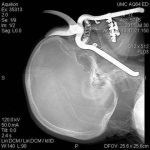
AAST 2011: Acute Kidney Injury And Multiple Organ Failure

Organ failure after major trauma is relatively common. Acute renal failure can occur for a variety of reasons, and tends to occur early. This abstract from Denver Health looked at acute kidney injury as a predictor for the development of multiple organ failure.

The authors retrospectively reviewed 12 years of their registry data for patients at high risk for developing organ failure. They found that multiple organ failure (MOF) developed in 21% and that 8% died. They also noted that if acute kidney injury (AKI, serum creatinine > 1.8mg/dL) occurred by day 2, it predicted the failure of additional organs. Specifically, 80% of these patients developed MOF, with a 34% mortality. Renal failure was a better predictor of multiple organ failure than heart, liver or pulmonary failure seen on day 2.

Bottom line: Early kidney failure, as shown by creatinine elevation, is a reliable predictor of multiple organ failure in severely injured patients. Prevention of acute kidney injury makes sense and may help, but further investigation is needed to demonstrate the mechanism.

Reference: Acute kidney injury and post-trauma multiple organ failure: the canary in the coal mine. AAST 2011 Annual Meeting, Paper 20.

AAST 2011: Preview Of The Annual Meeting

The 70th annual meeting of the American Association for the Surgery of Trauma begins on September 14 in Chicago. Starting tomorrow, I’m going to highlight some of the most interesting abstracts that are scheduled for presentation. Please recognize that I can only review the abstract itself, so my analyses will be limited. The complete manuscripts will not be available in published form for close to a year, and only if they are of a caliber to be accepted by the Journal of Trauma.

To download a pdf file containing all the oral abstracts, click here. For the poster abstracts, click here.

Return To Work After Severe Trauma

One of the most important goals after injury is return to work or school. There are some studies available that look at return to work/school status as a function of injury severity, demographic and insurance status. However, long-term studies are rare.

A Norwegian group followed a small population of injured patients very closely for five years, looking at the actual trajectory of return. They also tried to determine the specific factors that predicted return to work. The initial group numbered 101 people, but slowly decreased to 75 due to dropouts, nonresponders, and one patient who retired while receiving disability benefits.

The average age was 39 and ISS was 29. About 60% had a lower level of education and blue collar jobs. There were 28 patients with severe head injury, 12 with moderate head injury, 18 spinal cord injuries and 3 amputees among the group.

At the end of 5 years, only 49% had returned to work (see chart). 23% were on full disability and 9% on partial disability. Of greatest interest, there was only a small increase in return to work after 2 years. The best predictors of return to work were higher education level, good physical health and function (no surprise), and type of coping strategy. Time spent in rehab was also a factor.

Bottom line: Rehab that aims toward return to work is a major factor in getting better after major injury. However, an additional focus on coping and other psychological factors is important. Most people who will be capable of returning to work or school will do so by the two year mark.

Reference: Returning to work after severe multiple injuries: multidimensional functioning and the trajectory from injury to work at 5 years. J Trauma 71(2):425-434, 2011.

Brain Injury and DVT Prophylaxis Part II

I previously wrote about a new review that looked at using chemical prophylaxis for deep venous thrombosis (DVT) in patients with traumatic brain injury (TBI). The authors showed that it was safe to give subcutaneous heparin products within 24 to 48 hours after a stable 24 hour followup CT.

A just-published article now helps to refine the selection of the heparin product. A retrospective review looked at 386 ICU patients with a head Abbreviated Injury Score (AIS) > 2. A total of 57 received mechanical prophylaxis, the remainder received heparin products. Chemical prophylaxis consisted of subcutaneous enoxaparin 30mg bid or unfractionated heparin 5000u tid, at the whim of the attending neurosurgeon.

The heparin group had a slightly but significantly higher Head AIS (4.1 vs 3.8). The drugs were started at the same time post-injury, about 48 hours from admission. Unfractionated heparin was found to be inferior to enoxaparin. The unfractionated heparin patients had both a higher rate of pulmonary embolism, and were more likely to have progression of any intracranial hemorrhage (12% vs 5%). The authors claim a significantly lower DVT rate, but information in their data tables do not support this. Additionally, their overall DVT rate is very low, most likely because they did not routinely screen for it.

Bottom line: The head injury / DVT prophylaxis literature is expanding rapidly. It’s time to start working with your neurosurgeons to initiate chemoprophylaxis early (within 48 to 72 hours from injury once any intracranial bleeding is stable). And it looks like the drug of choice is enoxaparin, not unfractionated heparin.

Reference: Safety and efficacy of heparin or enoxaparin prophylaxis in blunt trauma patients with a head abbriviated injury severity score >2. J Trauma 71(2):396-400, 2011.

Related post: Brain injury and chemical prophylaxis for DVT

Technology: The VeinViewer

I’m always interested in technology that makes what we do easier. Here’s an objective look at an interesting machine that’s been around for a while. It uses near-infrared light to detect skin temperature changes to allow it to map out veins. It then projects an image of the map in real time onto the skin. In theory, this should make IV starts easier (as long as you can keep your head out of the way of the projector).

A paper just published from Providence, Rhode Island looked at this device to see if it could simplify IV starts in a tertiary pediatric ED. It was a prospective, randomized sample of 323 children from age 0 to 17 looking at time to IV placement, number of attempts, and pain scores.

Unfortunately, the authors did not find any differences. They found that nearly 80% of IVs were started on the first attempt with or without the VeinViewer, which is less than the literature reported 2-3 attempts. This is most likely due to the level of experience of the nurses in this pediatric ED. 

The authors did a planned subgroup analysis of the youngest patients (age 0-2) and found a modest decrease in IV start time (46 seconds) and the nurse’s perception of the child’s pain. Interestingly, the parents did not appreciate a difference in pain between the two groups. This may be due to the VeinViewer’s pretty green display acting as distraction therapy for the child.

Bottom line: This paper points out the importance of carefully reviewing all new (read: expensive at about $20,000 each) technology before blindly implementing it. In this case, an expensive peice of equipment can’t improve upon what an experienced ED nurse can already accomplish.

Reference: VeinViewer-assisted intravenous catheter placement in a pediatric emergency department. Acad Emerg Med, published online, doi: 10.1111/j.1553-2712.2011.01155.x, 2011.

I have no financial interest in Christie Digital Systems, distributor of the VeinViewer Vision®.

Home of the Trauma Professional's Blog

Do you want to get a daily email every time there’s a new post? See what I’m up to.

Click here to get details and subscribe!

[accua-form fid=”1″]

[mc4wp_form id=”2023″]