Temporal Bone Fracture

The temporal bone is a very complicated and important piece of bone located at the base of the skull. It houses a number of structures vital to hearing and balance, and is a conduit for several important nerves. Most fractures are due to blunt trauma, and although several classification systems exist, the majority are irregularly shaped and don’t lend themselves to easy categorization.

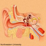
Clinically, a t-bone fracture should be suspected anytime that blood is seen in the ear canal or behind the eardrum after blunt head trauma. Patients may complain of dizziness or hearing loss. Examination may demonstrate some nystagmus in some cases. Most of the time, the diagnosis will not be made until a head CT is performed. The diagram above shows why this fracture can cause hearing problems (middle ear) or dizziness (inner ear).

There is usually no specific surgical intervention needed for this fracture. However, two important functional exams must be completed once diagnosed: facial nerve function and hearing. The facial nerve exam should document whether only portions of the nerve are affected vs all branches, and whether there is at least partial function. In head injured patients that can’t follow commands, even a grimace can demonstrate some function.

The ideal hearing test is carried out with a tuning fork. However, this is not usually convenient in acute trauma patients. Whispering and rubbing fingers near the patient’s ear can be used as a quick and dirty test. Awake patients will be able to tell you if their hearing is decreased.

Bottom line: Suspect temporal bone injuries in patients with any signs of basilar skull fracture. If the diagnosis is suspected or confirmed by CT scan, document the best facial nerve and hearing exam that you can. Contact your facial surgeon once the diagnosis is confirmed, and call them urgently if there is any loss of facial nerve function. Thin cut CT scans of the temporal bone are generally not necessary, and should not be ordered automatically unless the facial surgeon needs it for a specific reason.

Pneumothorax And Oxygen: The Final Post!

Okay, this is the last time I’m going to write about this. Hopefully I can provide the final nail in the coffin for this idea. Previously, the oldest paper I could find that was cited as a reason to use high inspired oxygen to treat pneumothorax was from 1983. I found what I think is the earliest (and the last that I will discuss) from 1971!

Twelve patients were retrospectively reviewed who recovered without intervention from a spontaneous pneumothorax. Another 10 were monitored prospectively with the same condition, but were given “high concentration oxygen” (??) by mask from 9 to 38 hours at a time. During intervening periods, the patients breathed room air. Daily chest xrays were obtained, and here is the cool part:

The inner edge of the chest wall and the outer edge of the lung were traced on transparent paper. This was then superimposed on graduated graph paper and the area corresponding to the pneumothorax cavity was measured. The rate of absorption was expressed in cm2/24 hrs.

Need I say more? The authors did show graphically that the apparent rate of absorption tripled in the treated patients, from about 5cm2/day to about 15cm2/day, and was higher in patients with a larger pneumothorax. The problem here is the same as before: chest xray does not allow volumetric estimates, so any results relying on them are suspect. At least it’s not a rabbit study.

Bottom line: There’s just no convincing data to support this practice, so let’s stop using it. Simple physics suggests that this should work, but the effect is just not clinically significant enough to offset the possibility of mishaps from an inpatient admission for oxygen therapy (see yesterday’s post). As I mentioned yesterday, look at the clinical status of your patient. If they have any detectable blood in their chest, they’ll probably need drainage. If not, and if they feel normal, discharge and follow up with a repeat xray in a week. The pneumo will probably be gone. If they do have some compromise, then insert the smallest tube you can. If done properly and a one-way valve can be used, the patient may still be managed as an outpatient.

Related posts:

Reference: Oxygen therapy for spontaneous pneumothorax. Br Med J 4:86-88, 9 Oct 1971.

More On Treating Pneumothorax With Oxygen

One of my readers has pointed out that, yes, the evidence for using O2 to treat pneumothorax is poor, but practice and standard of care are not always driven by evidence. He also pointed out that it’s not really fair to condemn the use of this modality if there isn’t specific evidence showing that it’s bad. In other words, doing something that seems benign is okay if we can’t show that it’s harmful or at least prove that it’s actually benign. I don’t agree.

My point is that no intervention is truly benign. There are always potential complications for the things we do as physicians, sometimes physical, sometimes psychological. Putting a patient on O2 seems safe. But if used as a treatment for pneumothorax, it means hospitalization (which costs a lot of money), an IV (which could get infected), exposure to a lot of sick people (read MRSA and other fun bugs), lying in bed a lot more than at home (DVT), and on and on.

If the pneumothorax does not interfere with function and the patient has decent pulmonary health, why not send them home with reassurance and get a followup chest xray at some point to confirm resolution? If it does cause physiologic problems, or they have pulmonary disease and are likely to develop complications such as pneumonia, then admit for the least invasive treatment to quickly get it out (pigtail type catheter).

Since this topic just won’t seem to die, I’m going to try to kill the last papers I’m aware of on this topic today and tomorrow. Today’s was published in a pediatric surgical journal (!), and it’s another rabbit study. This one adds a wrinkle to the one I discussed yesterday. Not only did they inject air to create a pneumothorax (20cc this time), they punctured the pleura with a needle to create an air leak to simulate a real clinical problem.

They saw the same trend as posted yesterday, although the times were longer. Once again, resolution was measured with chest xray (performed every 12 hours this time). Unfortunately, 7 of the 27 rabbits used in each group died, leaving only 6 or 7 in each of 3 groups for analysis (room air, 40%, 60% O2). Even with wide standard deviations, the authors claimed significant differences in recovery.

Same problems as yesterday, particularly with how resolution of pneumothorax is determined. And don’t use rabbits! A bigger issue is that this is not really a clinically relevant model. First, creating an air leak would defeat the overall purpose of giving high O2 concentrations. If 60% O2 leaked into the pleural space, there would be less nitrogen to wash out so one would think that resolution would take longer. And no one would consider treating a patient with an air leak without some type of drainage device for fear of a tension pneumothorax.

Bottom line: Still not enough evidence to support this seemingly benign treatment. Tomorrow I’ll look at the (hopefully) last paper on the topic since the beginning of time, published in 1971.

Related posts:

Reference: Supplemental oxygen improves resolution of injury-induced pneumothorax. J Pediatric Surg 35(6):998-1001, 2000.

Thanks to Jonathan St. George for his comments on yesterday’s post!

Treating Pneumothorax With Oxygen (Again)

The topic of treating pneumothorax with high inspired oxygen concentrations keeps coming up! I’ve written about this a few times in the past, and the literature I found supporting the practice was terrible. Some readers brought three more studies to my attention that support it, so I’m going to take the next three days to see if there is any hope for this practice.

Today’s paper used a rabbit model where each animal was given a complete pneumothorax by the injection of 15cc (!!) of air into one hemithorax. The authors then let the pneumothorax resolve using room air or 30%, 40%, or 50% FIO2. Each group consisted of 10 rabbits, and repeat chest xrays were obtained every 6-8 hours to follow resolution.

The statistical analysis was interesting and unusual. Because the authors were studying the time to resolved pneumothorax with higher inspired O2, they were looking for a test that would analyze an “ordered alternative.” The Jonckheere-Terpstra test was used, which I have never heard of, but I’ll assume it’s the legitimate one to use.

The figure at the top of this post shows the results. Looks promising right? There was a big improvement from room air to 30%, but lesser improvement using higher oxygen concentrations. The error bars (standard error of the mean) are remarkably tight, but this makes sense since xrays were only being taken every 6-8 hours.

The two big problems with this study are that: 1. they’re rabbits and it only takes 15cc of air to drop the entire lung, and 2. standard xray is being used to measure resolution. Trying to pick apart the exact time to resolution of a 15cc pneumothorax is very difficult, and to try to do it with a test that we know is not great at detecting small amounts of air even in big humans just doesn’t work. 

Bottom line: Fancy statistics and nice looking results don’t make up for an animal model that doesn’t necessarily correlate with humans and deriving results using an inaccurate diagnostic test. Tomorrow, I’ll look at a paper in the Journal of Pediatric Surgery to see if it fares any better.

Related posts:

Reference: Resolution of experimental pneumothorax in rabbits by graded oxygen therapy. J Trauma 45(2):333-334, 1998.

Thanks to Stephanie Taft MD at Regions Hospital for finding these fine studies for me.

Giving Rhogam (Rh Immunoglobulin) To A Man?

Rhogam is for women, right? The ATLS course points out that pregnant women who are Rh- and sustain significant blunt torso trauma should empirically receive Rhogam in case the fetus turns out to be Rh+.

But there is one situation where men might receive it. Most trauma centers use O- blood as their universal donor units because it does not contain any major antigens. However, O- blood is uncommon. Worldwide, only 4-9% of the population have this blood type. In China, the incidence of O- blood is nearly zero! So busy centers that don’t have much O- may substitute O+ blood for men. They then switch to the proper blood type when the crossmatch is complete

This makes sense, since men don’t ever have to worry about a Rh+ fetus. However, since this typically occurs at very busy (read: high penetrating injury) centers, there is a significant number of repeat offenders. And if they receive it again, the antibodies to the Rh factor they developed the first time can cause a significant hemolytic reaction. So men who receive O+ blood must be typed and given Rhogam if they are Rh-.

Reference: Emergency uncrossmatched transfusion effect on blood type alloantibodies. J Trauma 72(1):48-53, 2012.

Home of the Trauma Professional's Blog

Do you want to get a daily email every time there’s a new post? See what I’m up to.

Click here to get details and subscribe!

[accua-form fid=”1″]

[mc4wp_form id=”2023″]