All posts by The Trauma Pro

Trauma In Wine Country?

Nope. Sorry. The two don’t mix. I just finished attending the American College of Surgeons meeting in San Francisco, and now I’m moving up to the Napa Valley for a few days. However, enjoying the sights, food and wine here is not conducive to writing coherently.

So I’m taking a few days off to concentrate on other things. I’ll return on Tuesday with several days of material on field amputations. I’m also teaching several sessions at the TCAA Trauma Director course and their Trauma Performance Improvement course. I’ll be tweeting some of the more interesting content as I sit in on some of the others’ sessions.

Talk to you again on Tuesday. Cheers!

Emergency Care Of Bleeding From Dabigatran

Finally, a consensus report has been finalized by the Institute for Clinical Systems Improvement (ICSI) regarding bleeding in patients taking dabigatran (Pradaxa). I’ve written about the special problems posed by patients who are injured while taking this drug and related ones. I’ve also provided some management algorithms for consideration while complete ones were crafted. Well, here they are.

A workgroup of experts from hospitals here in Minnesota were convened to consider and provide a framework for managing these patients. A document was released recently to help guide their care.

To summarize, patients who experience a severe bleed, say from trauma, should be managed with:

  • Holding the medication
  • Evaluating bleeding. In trauma, this will generally involve CT scan.
  • Consider the need for surgery
  • Give activated charcoal if the drug was taken within 2 hours
  • Consider dialysis
  • Transfuse blood if hemoglobin / hematocrit needs to be improved
  • Infuse plasma after 4 units of red cells, and cryoprecipitate after 8 units packed cells / 4 units plasma if needed
  • Consider prothrombin complex concentrate or activated Factor VII in extreme cases

Click here to download the official document from ICSI.

Related posts:

What The Heck? The Answer

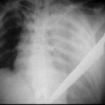
Obviously, there’s a big knife in this image, and it appears to coincide with our patient. Remember, you can’t tell the relative positions of objects in the z axis in a two dimensional radiograph. However, you know that it is not on top of or under this patient, because there is an obvious hemothorax.

First, this patient should have been a trauma activation, since there is a penetrating injury of the chest. The first decision point is based on vital signs: is the patient hemodynamically normal? If not, do the essentials quickly (quick exam, lines, blood draw for blood bank) and move to the OR. The quick exam is important, especially of the back, since you may not see it for the next few hours and additional injuries there could be trouble.

If the patient is stable, then it’s time to do the essentials and figure out what you are going to need to do in the next hour. A chest xray can be very helpful. However, given the size of the knife and the fact that it appears to be buried to the hilt allows you to calculate the trajectory in your mind. The only question is if it went in far enough to enter the abdomen.

The xray shows a moderate hemothorax and a tip that extends well into some part of the abdomen, no matter how you look at it. It has also come alarmingly close to the heart. If the knife had not penetrated as deeply and was obviously only in the chest, then removal of the knife with insertion of a chest tube would be appropriate. 

However, in the case of this xray, the patient still needs to go to the OR. The chest and abdomen need to be prepped and the abdomen opened first. Once the knife is located, the next steps can be planned. If there is concern for the heart, the pericardial surface of the diaphragm can be opened to look for blood. If found, extension of the laparotomy incision into a sternotomy is appropriate for cardiac repair. If not, the tip of the knife should be visualized and the weapon slowly removed under direct vision. Any bleeding structures can be dealt with first as it goes, followed by any injuries to a hollow viscus.

ED Thoracotomy Part 3: Clamping The Aorta

Finally, the chest is open and the tamponade has been relieved. But your patient has little volume. In order to conserve any circulating blood and pump it only to the heart and the head, it’s time to cross clamp the aorta. This task is best left to the surgeon, because it is not a simple matter.

First, you have to locate the aorta, ideally somewhere just above the diaphragm. Unfortunately, if the patient is hypovolemic it’s very difficult to distinguish the aorta from the esophagus, which lie right next to each other (see picture above). In order to make them feel different, insert a gastric tube through the mouth or nose.

Next, separate the aorta and esophagus. They are both covered by pleura. The structure nearest you without the tube in it will be the aorta. Sometimes it’s possible to use a finger to dissect through the pleura and around the aorta. However, the younger the patient, the tougher this tissue is. It may be necessary to incise the pleura with scissors while your assistant holds the lung anteriorly, our of the way.

Finally, once you can pass a finger completely around the aorta, use it to guide the placement of a gently curved DeBakey type clamp (see picture on the left). Squeeze it until it clicks once, and you are done! Now rapidly infuse warmed blood into the patient and run to the OR!

Related posts: