All posts by The Trauma Pro

Impact Of A Trauma Nurse Practitioner Model On Nonsurgical Admissions

Nonsurgical admissions are a concern for most verified/designated trauma centers. Under the current 2014 Resources for Optimal Care of the Injured Patient, all trauma patient admissions to a nonsurgical service must be concurrently reviewed by the trauma program. This process guards against trauma patients sneaking into the hospital on medicine services where the quality of the care for their injuries may not be monitored.

Typically, this requirement is met by having the trauma program manager (TPM), trauma PI coordinator (TPIC), or registrar run a daily admission report and mark patients with a potential trauma diagnosis for further review. Some clinician then reviews the patient in person or via a chart check. No further attention is needed if the patient has low acuity injuries or has been seen by the trauma or other surgical service. If not, additional scrutiny is recommended to identify patients who might be better off with a trauma service consult or even a transfer of service.

The trauma group at the Charleston Area Medical Center in West Virginia postulated that adopting a trauma nurse practitioner (TNP) model to provide care for patients otherwise admitted to a hospitalist service would improve care and decrease nonsurgical admissions. The nurses were supervised directly by the trauma attending surgeons.

They analyzed retrospective registry data during a 22-month period and compared nonsurgical admission rates with the same data for the preceding three years. Several performance metrics were evaluated, including length of stay and those statistics hospitals love to watch, discharge orders placed by noon, discharge location, and time to operating room.

Here are the factoids:

  • A total of 749 patients were admitted to the TNP service and 651 to hospitalist services
  • Patients in the TNP group were significantly younger (59 vs. 76) and more likely to be male
  • ISS was the same, but the TNP service patients had slightly more injuries (1.6 vs. 1.2) overall, and their patients were more likely to require an operation
  • The number of comorbidities was higher on the hospitalist service (2 vs. 1.6)
  • Hospital length of stay was one day shorter in the TNP group, which  was significant
  • TNP service patients were more likely to discharge home rather than to a skilled nursing facility
  • Time to OR for the TNP patients was significantly shorter by 11 hours
  • 30-day readmission rates were the same
  • The percentage of patients admitted to a nonsurgical service decreased from 20% to 14% after implementation

The authors concluded that the nonsurgical admission rate declined significantly, and several performance metrics also improved. In addition, the decreased length of stay was projected to result in a decrease of over $876,000 in hospital charges throughout the study.

Bottom line: This study illustrates some potential differences that surface when patients are admitted and managed by a trauma service rather than a hospitalist service. I say potential because there are a lot of confounders here.

The patients on the two services were very different. Although the TNP service patients had slightly more injuries (1.6 vs. 1.2), their ISS was identical. They also had fewer comorbidities. There appears to be some selection process in play here, but it was not explicitly stated in the paper. It also appears that older and more complicated patients, in terms of their comorbidities, were admitted to the hospitalists. Those with injuries more likely to require surgery were admitted to the TNP service.

Nonsurgical admission rates definitely decreased, but without knowing the selection criteria, this could have been due to just the presence of the TNP service and the desire to admit patients to it. The decreased hospital LOS and higher discharge rate to home are impressive, but could this also be due to these patients’ younger age?

The final issue is that the rules have changed! Starting later this year with the implementation of the 2022 Resource Document, the 10% nonsurgical admission rate threshold will disappear. Now, all nonsurgical trauma admissions must undergo primary review via the PI process. If any issues are identified, the ISS is greater than 9, or there was no trauma or surgical consultation, they must be escalated to a timely secondary review by the trauma medical director. Gone are the days of retrospective reviews of these cases!

What to do? It’s a balancing act in terms of trauma service capacity and staffing. Ideally, most injured patients are best served on the trauma or surgical specialty service. Several papers have outlined improved outcome metrics with this arrangement. Utilizing TNPs or physician assistants to capture and manage appropriate patients can definitely be helpful. 

An alternative is to integrate a hospitalist, preferably with geriatric expertise, into the trauma service so injured patients with more complex medical issues can be comfortably managed on the trauma service.

Reference: Rate of Nonsurgical Admissions at a Level 1 Trauma Center: Impact of a Trauma Nurse Practitioner Model. J Trauma Nurs 27(3):163-169, 2020.

 

PAs and NPs In Level I Trauma Centers

Trauma service staffing is important to maintaining trauma center status. Teaching centers in the US have been grappling with resident work hour rules, and non-teaching centers have always had to deal with how to adequately staff their trauma service. What is the impact of staffing a trauma center with midlevel practitioners (MLPs) such as physician assistants and nurse practitioners?

A state designated Level I trauma center in Pennsylvania retrospectively examined the effect of adding MLPs to an existing complement of residents on their trauma service. They examined the usual outcomes, including complications, lengths of stay, ED dwell times and mortality.

Here are the more interesting factoids:

  • ED dwell time decreased for trauma activations and transfers in, but it increased for trauma consults. Of note, data on dwell times suffered from inconsistent charting.
  • ICU length of stay decreased significantly
  • Hospital length of stay decreased somewhat but did not achieve significance
  • The incidence of most complications stayed the same, but urinary tract infection decreased significantly
  • There was no change in mortality

Bottom line: There is a growing body of literature showing the benefits of employing midlevel providers in trauma programs. Whereas residents may have a variable interest in the trauma service based on their career goals, MLPs are professionally dedicated to this task. This study demonstrates a creative and safe solution for managing daily clinical activity on a busy trauma service.

In my next post, I’ll review a more recent paper gauging the impact of a trauma nurse practitioner model on managing non-surgical admissions.

Reference: Utilization of PAs and NPs at a level I trauma center: effects on outcomes. J Amer Acad Physician Assts, July 2011.

Diagnosing BCVI In Children

Several days ago, in my post on “How Common Is BCVI?” I mentioned a paper recognizing the increasing incidence of BCVI in pediatric patients and the very high stroke rate (37%) and death rate (13%). These numbers are very concerning!

Previous work shows that the Memphis and Denver criteria are not very sensitive in adults. This has led many trauma centers to add CT angiography of the neck automatically in patients with a high-energy mechanism. But what about kids? Are these screening criteria any better?

A consortium of children’s hospital trauma centers has a paper currently in press that evaluated both the Memphis and Denver criteria in children under age 15. It was a four-year prospective, observational study of children with head, face, or neck injuries. Although the Memphis criteria were specifically used in the study, data for applying the original and expanded Denver criteria, EAST practice management guideline criteria, Utah score, and McGovern score were also collected. The last two are pediatric-specific criteria.

Any child who met at least one of the Memphis criteria received a CTA or MRA of the head and neck. In addition, all children with head, face, or neck injuries received a follow-up evaluation two weeks after discharge. This was designed to capture any evidence of BCVI in those who did not meet Memphis criteria and hence had no CTA/MRA. Patients who missed this evaluation or had other missing data were excluded from the analysis.

Here are the factoids:

  • A total of 2,284 children met the criteria for enrollment; nearly one-third were excluded due to no imaging/follow-up or missing data (!)
  • There were 24 BCVI diagnosed (1.6%)
  • Diagnostic accuracy of the various screening criteria were:
Criteria Sens Spec PPV NPV # CTA to detect one BCVI
Memphis 92 71 5 100 20
Denver 73 88 9 100 11
Expanded Denver 88 64 4 100 25
EAST 79 83 7 100 14
Utah 49 96 16 99 7
McGovern 75 90 11 100 9

The Memphis criteria had the highest sensitivity and would have missed the fewest BCVI. The pediatric-specific Utah score had the highest specificity but would have missed more than half of the injuries. The authors recommend refining the Memphis criteria to improve its specificity while maintaining its high sensitivity.

Bottom line: As with adults, we struggle with systematically identifying BCVI. All screening systems leave something to be desired. It’s not practical or prudent to treat children the same as adults and just liberalize the use of CTA. Substituting MRA is not practical because this requires sedation and/or intubation in the younger age groups.

Of interest in this study, the overall incidence was higher (1.6% here vs. less than 0.5% in my previous post). This is probably due to the fact that there was a significant effort to identify criteria for angiography, and follow-up was provided to detect occult injuries.

This paper adds to the previous work I cited describing how important it is to detect this injury. The current research demonstrates that the Memphis criteria are the best we have for pediatric patients at this time. But it clearly shows the need for a better tool. 

But until one is developed, a best practice would be to use the Memphis criteria to screen any pediatric patient with head, neck, or facial trauma due to a high-energy blunt mechanism. Then select CTA or MRA after conferring with your pediatrics and radiology teams.

Reference: Diagnostic accuracy of screening tools for pediatric blunt cerebrovascular injury: An ATOMAC multicenter study. Journal of Trauma, publish ahead of print, DOI: 10.1097/TA.0000000000003888.


Treatment Of BCVI

In my last post, I reviewed the grading system for blunt carotid and vertebral artery injury (BCVI). Today, we’ll discuss treatment, and in the next post, we will wrap up with pediatric-specific information.

There are basically three modalities at our disposal for managing BCVIantithrombotic medication (heparin and/or antiplatelet agents), surgery, and endovascular procedures. The choice of therapy is usually based on surgical accessibility and patient safety for anticoagulation. We know that several studies have shown decreased stroke events in heparinized patients. Unfortunately, this is not always possible due to associated injuries. Antiplatelet agents are usually tolerated after acute trauma, especially low-dose aspirin. However, several studies have shown little difference in outcomes in patients receiving heparin vs. aspirin/clopidogrel for BCVI.

So what to do? Here are some broad guidelines:

  • Grade I (intimal flap). Heparin or antiplatelet agents should be given. If heparin can be safely administered, it may be preferable in patients needing other surgical procedures since it can be rapidly reversed by stopping the infusion. These lesions generally heal entirely on their own, so a follow-up CT angiogram should be scheduled in 1-2 weeks. Medication can be stopped when the lesion heals.
  • Grade II (flap/dissection/hematoma). These injuries are more likely to progress, so heparin is preferred if it can be safely given. Stenting should be considered, especially if the lesion progresses. Long-term anti-platelet medication may be required.
  • Grade III (pseudoaneurysm). Initial heparin therapy is preferred unless contraindicated. Stable pseudoaneurysms should be followed with CTA every six months. If the lesion enlarges, surgical repair should be performed in accessible injuries or stenting in inaccessible ones.
  • Grade IV (occlusion). Heparin therapy should be initiated unless contraindicated. Patients who do not suffer a catastrophic stroke may do well with follow-up antithrombotic therapy. Endovascular treatment does not appear to be helpful.
  • Grade V (transection with extravasation). This lesion is frequently fatal, and the bleeding must be addressed using the best available technique. For lesions that are surgically accessible, the patient should undergo the appropriate vascular procedure. Inaccessible injuries should undergo angiographic treatment and may require embolization to control bleeding without regard for the possibility of stroke.

References:

  1. Scott WW, Sharp S, Figueroa SA, et al. Clinical and radiographic outcomes following traumatic Grade 1 and 2 carotid artery injuries: a 10-year retrospective analysis from a Level I trauma center. J Neurosurg 122:1196, 2015.
  2. Scott WW, Sharp S, Figueroa SA, et al. Clinical and radiographic outcomes following traumatic Grade 3 and 4 carotid artery injuries: a 10-year retrospective analysis from a Level 1 trauma center. J Neurosurg 122:610, 2015.
  3. Scott WW, Sharp S, Figueroa SA, et al. Clinical and radiological outcomes following traumatic Grade 1 and 2 vertebral artery injuries: a 10-year retrospective analysis from a Level 1 trauma center. J Neurosurg 121:450, 2015.
  4. Scott WW, Sharp S, Figueroa SA, et al. Clinical and radiological outcomes following traumatic Grade 3 and 4 vertebral artery injuries: a 10-year retrospective analysis from a Level I trauma center. The Parkland Carotid and Vertebral Artery Injury Survey. J Neurosurg 122:1202, 2015.

How To Grade BCVI

In my last post, I reviewed the three screening systems for blunt carotid and vertebral artery injury (BCVI). Today, we’ll look at grading them.

Just ten years ago, it was a major production to identify BCVI. Then, CT angiography was still in its early days, and scanner resolution and radiologist experience were significant issues that impacted accuracy.

We’ve come a long way in a relatively short period of time, and current-day scanners now have more than adequate resolution. It’s also more common to have a radiologist with special skills reading these studies, the neuroradiologist. For these reasons, CT angiography has become the standard for diagnosis. It is also the most cost-effective. Only in sporadic cases do we need to obtain a conventional contrast angiogram.

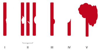
Once the study has been obtained, it’s time to identify and classify the injury. The Denver group is also responsible for bringing us the grading system for BCVI. See the diagram below.

Here are the details:

Grade I: A mild intimal irregularity is seen. Note the abnormally narrowed area, representing a minor intimal injury, possibly with a small amount of clot.

Grade II: This grade has several presentations. There may be an intraluminal thrombosis/hematoma with (left) or without (right) an intimal flap, or a flap alone (center).

Grade III: There is a full-thickness injury to the vessel with a contained extraluminal extravasation (pseudoaneurysm)

Grade IV: The vessel is completely occluded by a flap or thrombus

Grade V: The artery is transected and freely extravasating

Here’s a nice diagram:

Remember, we always grade things for a reason! Ultimately, the injury grade will translate into the selection of treatment. We’ll cover that in my next post.

Reference: Blunt carotid arterial injuries: implications of a new grading scale. J Trauma. 1999;47(5): 845-53.