All posts by The Trauma Pro

The Poor Man’s Water Seal Test

Placing the chest tube collection system to water seal can be a valuable tool in determining if it’s time to remove the tube. The idea is that removing suction from the system will help identify a slow air leak that may not be obvious while watching the collection system. Typically, water seal without suction is maintained for 6 hours and a chest xray is obtained to look for a new (or larger) pneumothorax.

image

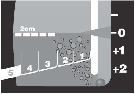
Here’s a way to detect a small air leak faster. When you enter the patient room, disconnect the suction tubing from the collection system. Chat with your patient, and do your usual thorough exam. Take your time. When you are done, slowly slip the suction tubing back onto the collection system. Watch the water seal chamber closely for any bubbles that try to sneak under the partition (above).

If the fluid level slowly lowers, but no bubbles pass the partition, there is no significant leak. On occasion, I’ve seen a single bubble creep under, and this is probably okay. However, if a train of bubbles passes, an air leak is present and it is premature to consider pulling the tube. Place the system back on suction.

Like so many tests, this one is only helpful if a leak is seen. It means that a significant amount of air has accumulated in the short time you’ve been examining your patient. If there are no bubbles, there still could be a very slow leak, so they will still need a formal water seal test, if indicated. See the protocol below for details.

Related post:

What The Heck? Bicycle Trauma Answer

So a young male jammed a handlebar into his abdomen, and a CT image demonstrating his problem was shown. But what did it actually show?

By now, you probably realize that clinical information is key. On exam, he had an obvious bulge in his left lower quadrant, more obvious with straining. Looking at the CT (now with a nice arrow), there is a problem over the left side of the abdomen. 

image

This child has so little fat, that it’s difficult to see the problem. If you track the thin layer of fat across the abdomen to the right side of the image, you’ll see that it disappears over the bowel gas. This represents a complete tear through all fascial layers, not just a Spigelian hernia as some readers guessed.

Management consisted of primary repair of the defect. An uneventful recovery can be expected. Unless more bicycle tricks are anticipated.

Reference: Traumatic handlebar hernia: a rare abdominal wall hernia. J Ped Surg 39(10):e20-e22, 2004.

What The Heck? Bicycle Trauma Part 2

Yesterday’s puzzle involved a young male who drove a handlebar into his abdomen. Little additional information was given, other than one slice of his abdominal CT scan. So what’s the problem?

The textbooks always associate handlebars with pancreatic and duodenal injuries, and these should always be looked for. However, the scan slice in this case was taken lower, within the pelvis. Too low to show you either of those organs.

As I’ve said before, be systematic when reading xray images. We automatically focus on the viscera and bones. Look at those areas, make sure you can identify each structure that you see, and look for any anomalies. 

But don’t forget the soft tissue! In this case, the child doesn’t have much. Take a closer look at the same slice and see if you can figure it out by tomorrow.

ICP Monitoring: Less Is More?

Management of severe traumatic brain injury (TBI) routinely involves monitoring and control of cerebral perfusion pressure. Monitoring is typically accomplished with an invasive monitor, with the extraventricular drain (EVD) and fiberoptic intraparenchymal monitors (IP) being the most common.

The extraventricular drain is preferred in many centers because it not only monitors pressures, but it can also be used to drain cerebrospinal fluid (CSF) to actively try to decrease intracranial pressure (ICP). But could less really be more? Surgeons at Massachusetts General reviewed 229 patients with one of these monitors, looking at outcomes and complications. They found the following interesting results:

  • There was no difference in mortality between the two monitor types
  • The EVD patients did not require surgical decompression as often, possibly because of the ability to decrease ICP through drainage
  • The EVD patients were monitored longer, and had a longer ICU length of stay. This was also associated with a longer hospital length of stay.
  • Complications were much more common in the extraventricular drain group (31%). The most common complications were no drainage / thrombosis (15%) and malposition (10%). Hemorrhage only occurred in 1.6% of patients. 
  • Fiberoptic monitors had a lower complication rate (8%). The most common was malfunction leading to loss of monitoring (12%). Hemorrhage only occurred in 0.6% of patients.

Bottom line: Don’t change your monitoring technique yet. Much more work needs to be done to flesh out this small retrospective study. But it should prompt us to take a critical look for better indications and contraindications for each type of monitor.

Reference: Intraparenchymal versus extracranial ventricular drain intracranial pressure monitors in traumatic brain injury: less is more?J Am Coll Surg 214(6):950-957, 2012.