All posts by The Trauma Pro

Could Be A Urethral Injury, But The Catheter’s Already In?

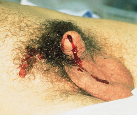
You’re seeing a trauma patient, probably a transfer from somewhere else. Either they told you there “may have been” some blood at the tip of the urethra, or maybe you see it smearing the outside of a urinary catheter that’s already in place! How do you proceed from here?

First, try not to get into that situation. Make sure that everyone on your team knows that gross blood at the meatus, male or female, means urethral injury until proven otherwise. If it’s not gross blood, it could be that the patient was incontinent and has hematuria from other causes. The fear with passing a catheter across a urethral injury is that it may convert a partial tear to a complete one. Reconstruction and complications from the latter are far more serious.

But the catheter is there. What to do?

First, leave the catheter in place. You must assume that the injury is present, and you need to rule it in or rule it out in order to decide what to do with the catheter. If the injury is not really there, then you can remove the catheter when indicated. If it really is present, then the urethral injury is being treated appropriately.

Next, do a urethrogram. I’ve previously described how to do it here, but the technique I describe is only appropriate for uncatheterized patients. The technique must be modified to use thin contrast and a method to inject alongside the catheter. To do this, fill a 20-30cc syringe with contrast (Ultravist or similar liquid) and put an 18 gauge IV catheter on the tip (no needles, please). Slide the IV catheter alongside the urinary catheter, clamp the meatus with your fingers, pull the penis to the side and inject under fluoroscopy. The contrast column will not be as vivid as with a regular urethrogram because it is outlining the urinary catheter, so there is less volume.

If the contrast travels the length of the urethra and enters the bladder without leaking out into soft tissue, there is no injury. If there is contrast leakage, stop injecting and plan to call a urologist.

Finally, be on the lookout for associated injuries. Urethral injuries are frequently found in patients with anterior pelvic fractures and perineal injuries.

Related post:

Link: blood at the urethral meatus (Atlas-Emerg-Medicine.org.ua from McGraw-Hill)

Thoughts On Traumatic Hematuria: Part 2

Yesterday, I discussed blood in the urine from a urethra. As I mentioned, there is typically not much from that particular injury. Today, I’ll dig into the three causes of real hematuria.

All of these tubes show gross hematuria except the one on the right.

  • Bladder injury. This can occur with either blunt or penetrating injury. The degree of hematuria is variable with stabs or gunshots, but tends to be much darker in blunt injury. This happens because the size of the bladder injury tends to be greater with blunt force. The bladder injury is not necessarily full-thickness with blunt trauma. It may just be some wall contusion and underlying mucosal injury. But frequently, with seat belt injury and/or A-P compression injuries to the pelvis (“open book”), the injury is full thickness.
    • Tip: If less than 50cc of very dark urine flow from the catheter upon insertion, it is likely that your patient has an intraperitoneal bladder rupture!
  • Ureteral injury. This injury is very rare. The most common mechanism is penetrating, but this structure is so small and deep that it seldom gets hit by naything. Patients with multiple lumbar transverse process fractures will occasionally have a small amount of hematuria, probably from a minor contusion. More often than not, the hematuria is microscopic, so we should never know about it.
  • Kidney injury. The most important fact regarding renal injury is that the degree of injury has no correlation with the amount of hematuria. The most devastating injury, a devascularized kidney, frequently has little if any gross hematuria. And conversely, a very minor contusion can produce very red urine.

So what about diagnosis? It’s easy! If you see gross hematuria, insert a foley catheter (if not already done) and order a CT of the abdomen/pelvis with contrast, as well as a CT cystogram. The latter must not be done using passive filling of the bladder with a clamped catheter. Contrast must be infused into the bladder under pressure to ensure a bladder injury can be identified.

CT scan is an excellent tool for defining injuries to kidney, ureter, and bladder, and will identify extravasation into specific places and allow grading. Specific management will be the topic of future posts.

Thoughts On Traumatic Hematuria: Part 1

I’ve seen a number of patients recently with bloody urine, and that is prompting me to provide some (written) clarity to others who need to manage this clinical problem. I’ll try to keep it organized!

There are two kinds of hematuria in trauma: blood that you can see with the naked eye, and…

Okay, so there’s only one. Trauma professionals do not care about microscopic hematuria. It does not change clinical management. Sure, your patient might have a renal contusion, but you won’t do anything about that. Or, he/she might have an infarcting kidney. And you can’t do anything about that. If you order a urinalysis, you might see a few RBCs. Don’t let this lead you down the path of looking for a source. You’ll end up ordering lots of tests and additional imaging, and generally will have nothing to show for it at the end. It’s not your job to spend good money on the very rare chance of finding something clinically significant.

Both of these specimens have blood in them. You can’t see it on the left, so don’t go looking for it with a microscope.

There are four sources of blood in the urine.

1. The first source does not generally cause hematuria, but can occasionally cause a few visible wisps of blood. That source is a urethral injury. The textbook teaching, and it’s good advice, is to look at the urethral meatus in your trauma patient, especially if you are contemplating insertion of a urinary catheter. If you see a few drops of blood, pause to consider. Sometimes, the blood is no longer visible, but might be present as a few well-placed drops on the patient’s underwear. So have a look at that, too, especially in patients with high risk injuries such as A-P compression pelvic fractures (think, lots of ramus fractures or pubic diastasis).

If you didn’t notice it and inserted the catheter anyway, you might see a few wisps of blood in the tubing as you place it. More often than not, this is just run of the mill irritation of the mucosa by the catheter, but always keep the possibility of an injury in mind.

Tomorrow, I’ll discuss the remaining three sources, and what to do about them.

Related posts:

The August Trauma MedEd Newsletter Is Coming Soon: The Laws of Trauma

I’m going to send out the next edition of the Trauma MedEd newsletter early next week. In this one, I’ll be presenting and discussing some of the “Laws of Trauma” that I’ve observed over the years. I think you’ll find them interesting and amusing.

As always, this issue will go to all of my subscribers first. If you are not yet one of them, click this link to sign up and/or download back issues.

Unfortunately, non-subscribers will have to wait until I release the issue on this blog, about 10 days later. So sign up now!

When Can You Take A Hypotensive Patient To CT?

The last two posts, I went on a rant about taking hypotensive patients to CT. The bottom line is that this is a generally bad idea, even if bad papers say it’s okay. However, we all know that there are no absolutes, especially in trauma.

So yes, there are two cases where one could justify taking a hypotensive patient to CT scan. Here they are:

  1. You believe that your patient has a catastrophic brain injury which is responsible for the hypotension. You would like CT confirmation so you can begin to withdraw support and terminate any other interventions.
  2. Your patient has sustained a cervical spinal cord injury and has neurogenic shock. You have started fluid resuscitation and are considering a pressor to normalize blood pressure, but would like to continue your diagnostic routine.

But before you can even consider leaving your resuscitation room, you must ensure that there is no other source of hypotension. This means getting chest and pelvic xrays to look for hemothorax or fractures. It means getting a good FAST exam to make sure there is no significant hemoperitoneum. It also means making sure that any fractures are properly splinted and there is no uncontrolled external bleeding.

You can only go to CT scan once all of these other potential bleeding sources have been ruled out. If in doubt, you must proceed to OR to either stop the bleeding or prove that it does not exist.

Are there any other reasons to take one of these patients to CT that you can think of? If so, leave comments or tweet!

Related post: