Blunt Carotid And Vertebral Artery Injury (BCVI): First Adults, And Now In Children!

I’ve written quite a bit about the challenges of diagnosing blunt carotid and vertebral artery injury (BCVI) in adults.  And now some papers on the potential danger to pediatric patients are beginning to surface.

I think it’s time to repost and update my series on BCVI. I’ll start with the basics, like nomenclature. I’ll then move on to how often we actually encounter it. This will include new information on younger patients.

I’ll dig into the various screening systems, and will include new information from some recent pediatric papers. The, I’ll finish up with how to grade it and suggested treatment routines based on grade.

Lets start with the acronym itself. There seems to be some confusion as to what BCVI actually stands for. Some people believe that it means blunt cerebrovascular injury. This is not correct, because that term refers to injury to just about any vessel inside the skull.

The correct interpretation is blunt carotid and vertebral artery injury. This term refers to any portion and any combination of injury to those two pairs of vessels, from where they arise on the great vessels, all the way up into the base of the skull. Here’s a nice diagram:

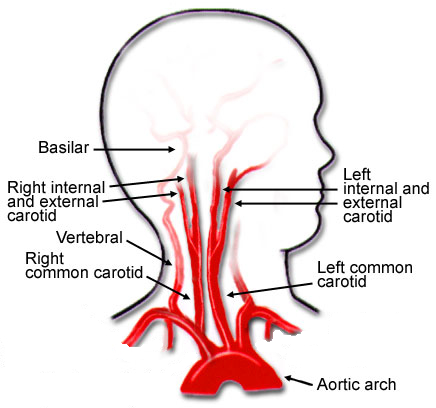
Note that we will be excluding the external carotid arteries from this discussion, since injuries to them do not have any impact on the brain. They can cause troublesome bleeding, though.

These arteries are relatively protected from harm during blunt trauma. But given enough energy, bad things can happen. Fortunately, injuries to these structures are not very common, but unfortunately many trauma professionals under-appreciate their frequency and severity.

In the next post, we’ll explore the incidence of this injury in both adults and children. Is it truly as uncommon as we think?

Subdural Hematomas and Hygromas Simplified

There’s a lot of confusion about subdural pathology after head trauma. All subdural collections are located under the dura, on the brain’s surface. In some way, they involve or can involve the bridging veins, which are somewhat fragile and get more so with age.

Head trauma causes a subdural hematoma by tearing some of these bridging veins. Notice how thick the dura is and how delicate the bridging veins are in the image below.

When these veins tear, bleeding ensues, which layers out over the surface of the brain in that area. If the bleeding does not stop, pressure builds and compresses and shifts the brain. A subdural hematoma is considered acute from the time of injury until about three days later. During this time, it appears more dense than brain tissue.

After about 3-7 days, the clot begins to liquefy and becomes less dense on CT. Many hematomas are reabsorbed, but occasionally there is repeated bleeding from the bridging veins, or the hematoma draws fluid into itself due to the concentration gradient. As a result, it can enlarge and begin to cause new symptoms. During this period, it is considered subacute.

It moves on to a more chronic stage over the ensuing weeks. The blood cells in it break down completely, and the fluid that is left is generally less dense than the brain underneath it. The image below shows a chronic subdural (arrows).

Hygromas are different because they are a collection of CSF, not blood. They are caused by a tear in the meninges and allow CSF to accumulate in the subdural space. This can also be caused by head trauma and is generally very slow to form. They can lead to slow neurologic deterioration and are often found on head CT in patients with a history of falls, sometimes in the distant past. CT appearance is similar to a chronic subdural, but the density is the same as CSF, so it should have the same appearance as the fluid in the ventricle on CT.

The January Trauma MedEd Newsletter Is Available!

The January issue of the Trauma MedEd newsletter is now available to everyone!

This issue contains a collection of miscellaneous interesting stuff.

In this issue, you will learn about:

  • Nonsurgical Admissions And The Nelson Score
  • Tip: Evaluation of Hematuria in Blunt Trauma
  • Central Lines Cause Hypercoagulability?
  • Lab Values From Intraosseous Blood
  • Leukocytosis After Splenic Injury

To download the current issue, just click here! 

Or copy this link into your browser:  https://www.traumameded.com/courses/interesting-stuff-1/

This newsletter was released to subscribers a week ago. If you would like to be the first to get your hands on future newsletters, just click here to subscribe!