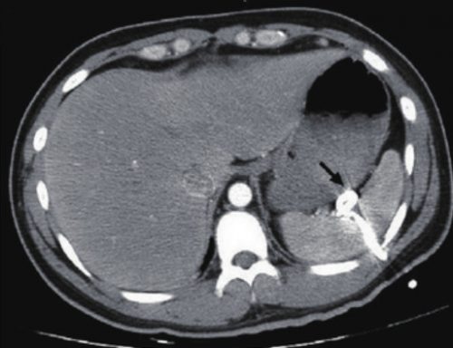
Here’s a case to make you think!
A patient arrives after being t-boned in his driver side door. He complains of left sided chest and abdominal pain. Chest x-ray shows a modest left hemopneumothorax. The decision is made to insert a pigtail type chest tube, and this is carried out in your trauma bay. It is uneventful, and a small amount of blood but no air is returned. The pelvis x-ray is unremarkable
The patient is then taken to CT, where an abdomen/pelvis scan with contrast is performed. This interesting slice is noted. What the heck?!

Here are my questions:
- What is wrong in this picture?
- How could it have been avoided?
- Does a pigtail chest tube work for hemothorax?
- How should this issue be managed, and where?
I’ll address these questions in my next post, and more!
Image source: internet

