How To: Manage Extraperitoneal Bladder Rupture

Extraperitoneal bladder rupture is a relatively uncommon injury, but is easily managed in most cases. It is associated with a blunt mechanism, and concomitant fracture of the pubic rami or spreading of the symphysis pubis is nearly always present. In the old days, we used to think that the bladder injury was due to penetration anteriorly by bony fragments, but this is probably an old wives tale. It’s more likely due to hydraulic forces occurring within the bladder at the same time the pelvic ring is being deformed or spread apart by blunt forces.

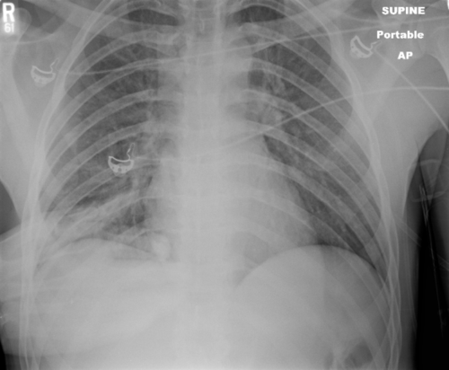
If you obtain a pelvic x-ray during the initial trauma evaluation and see any fractures or diastasis around the symphysis, think bladder injury. Placement of a urinary catheter will typically drain plenty of urine, which will usually be grossly bloody.

Once the injury is suspected, the diagnostic test of choice is a CT cystogram. Don’t confuse this with the images seen when the bladder passively fills with contrast when the catheter is clamped. There is not enough pressure in the bladder to guarantee that contrast will leak out, so this type of study may be falsely negative.

True CT cystogram technique requires filling the bladder with at least 350cc of dilute contrast under pressure by hanging it on an IV pole, then clamping the catheter. Once the bladder is filled, the scan can proceed as usual. But after it is complete, a second limited scan through the pelvis must be performed after the contrast has been evacuated by unclamping the catheter. This allows visualization of small contrast leaks that might otherwise be masked by all the contrast in the bladder.

Here’s a nice sagittal image of an extraperitoneal injury from radiologypics.com:

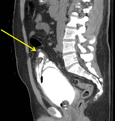
Note how the contrast dissects around the bladder but does not enter the peritoneal cavity.

Extraperitoneal injuries usually do not require repair and will heal on their own. However, if the symphysis pubis needs instrumentation to restore anatomic position, concomitant repair of the bladder is frequently necessary to keep the hardware from being contaminated by urine.

Bottom line:

  • Suspect an extraperitoneal bladder injury in anyone with bony injuries involving the symphysis pubis.
  • Don’t order a urinalysis in trauma patients!
  • Use CT cystogram technique to make the diagnosis.
  • Treatment is simple: leave the urinary catheter in place for 10 days. No urology consult is needed.
  • Then repeat the CT cystogram to confirm healing, and remove the catheter.

Related posts:

ABI vs API For Vascular Trauma

In general, the first maneuver in evaluating for possible vascular injury in an extremity is the good old physical exam. Is there a pulse or isn’t there? You can then subdivide that into: is the pulse weaker than normal. The problem is, what is “normal?” In most cases, we just compare it to another pulse somewhere and make a subjective judgment.

But we love to be more objective about things. Over the years, two simple, noninvasive techniques for evaluating pulses have been developed. The first is the ankle brachial index (ABI) , which was first described in 1930 and was used for diagnosis of peripheral vascular disease in 1950. It is performed by dividing the systolic pressure at the ankle of the affected extremity by the systolic pressure of one of the brachial arteries in the arms.

The new(er) kid on the block is the arterial pressure index (API), first described in 1991. This value is calculated by dividing the systolic pressure in the affected extremity by the systolic pressure in the contralateral uninjured extremity.

Many trauma professionals use the ABI when evaluating for potential vascular trauma. The typical threshold for pursuing further evaluation is 0.9, and several papers have been published on this topic. The API has also been critically evaluated, and the same threshold is used.

However, I believe that the API is more relevant and accurate than ABI. Why? Patients with atherosclerotic disease typically manifest it in their lower extremities. This serves to falsely elevate the ABI to a value greater than 1.0. It becomes more difficult to get down to that critical value of 0.9 that might indicate a vascular injury. Thus, the ABI may not detect a true injury, especially one in the lower extremities.

The API, on the other hand, relies on the fact that the amount of atherosclerotic disease is usually symmetric between the two lower extremities or the two upper extremities. Thus, the value will not be falsely elevated and will more accurately reflect the presence or absence of a vascular injury.

Bottom line: I recommend that you use the API when evaluating extremity vascular injury. Calculate the ratio by dividing the systolic pressure in the injured extremity with the pressure in the contralateral uninjured extremity (if there is one). A value < 0.9 indicates the need for angiographic evaluation, usually by CT scan.

And here’s a nice algorithm for managing peripheral vascular trauma from Life in the Fastlane:

Reference: Can Doppler Pressure Measurement Replace “Exclusion” Arteriography in the Diagnosis of Occult Extremity Arterial Trauma? Ann Surg 214(6):737-741, 1991.

Don’t Repeat Trauma Activation X-rays!!

You are in the middle of a fast-paced trauma activation. The patient is awake, and mostly cooperative. The x-ray plate is under the patient and everyone stands back as the tech gets ready to fire the x-ray machine. At that very moment, your patient reaches up and places his hand on his chest. Or one of the nurses reaches over to check an IV site.

The x-ray tech swears, and offers to re-shoot the image. What do you do? Is it really ruined? They have an extra plate in hand and are ready to slide it under the patient bed.

The decision tree on this one is very simple. There are two factors in play: what do you need to see, and how hard is it to see? The natural reaction is to discard the original image and immediately get a new one. It’s so easy! Plus, the techs will take heat from the radiologist because of the suboptimal image. But take a look at this example of a “ruined” chest xray.

It’s just the patient’s hand! You can still see everything that you really need to.

Bottom line: You are looking for 2 main things on the chest x-ray: big air and big blood. Only those will change your management in the trauma bay. And they are very easy to see. Couple that with the fact that an arm overlying the image does not add a lot of “noise” to the image. So look at the processed image first. 99% of the time, you can see what you need, and will almost never have to repeat. [Hint: the same holds true for the pelvic x-ray, too. You are mainly looking for significant bony displacements, which are also easy to see.]

Related posts:

Spleen Embolization In Adolescents?

Modern day nonoperative management of solid organ injury in adults came to be due to its success rate in children. But if you look at the practice guidelines for adults, they frequently include a path for angioembolization in certain patients. In children, embolization is almost never recommended.

But what about that gray zone where children transition to adults? How young is too young to embolize? Or how old is too old not to consider it?

The adult and pediatric trauma groups at Wake Forest looked at this question by reviewing their respective trauma registry data. They looked specifically at patients age 13-18 who presented with a blunt splenic injury over a 8.5 year period. About halfway through this period, adult patients (> 16 years) were sent for embolization not only for pseudoaneurysm or extravasation, but also for high grade injury (> grade 3).  Patients under age 16 were managed by the pediatric trauma team, and those 16 and older by the adult team.

Here are the factoids:

  • Of the 133 patients studied, 59 were “adolescents” (age 13-15) and 74 were “adults” (16 or older)
  • Patients managed by the adult team sent 27 of their 74 patients for angiography
  • Those managed by the pediatric team were never sent to angiography
  • The failure rate for nonoperative management was statistically identical, about 4% in adults and 0% in adolescents
  • For high grade injuries, the adult team sent 27 of 34 patients to IR, whereas the pediatric team sent none of 36. Once again, failure rate was identical.

Bottom line: We already know that too many adult trauma centers send too many younger patients to angiography for solid organ injury. This study tries to tease out when a child becomes an adult, and therefore when angiography should begin to be considered. And basically, it showed that through age 15, they can still be considered as and treated like children, without angiography.

But remember, these numbers are relatively small, so take this work with a grain of salt. If you are managing a younger patient nonoperatively, and they continue to show evidence of blood loss (ongoing fluid/blood requirements, increasing heart rate), angiography may be helpful in avoiding laparotomy as long as your patient remains hemodynamically stable. But consult with your friendly neighborhood pediatric surgeon first.

Related posts:

Reference: The Spleen Not Taken: Differences in management and outcomes of blunt splenic injuries in teenagers cared for by adult and pediatric trauma teams in a single institution. J Trauma, in press, May 2017.