Can You Teach A Trauma Surgeon To Insert An ICP Monitor?

You’ve heard the statistics about the graying of our society. The proportion of older people is growing rapidly. Well, there are only about 4400 neurosurgeons in the US, and they are aging as well. Nearly a third are older than 55 years.

This leaves a relatively small number of neurosurgeons tasked with helping to take care of trauma patients. Many Level II centers are hard pressed to maintain their neurotrauma services. Even basic procedures like ICP monitor placement may require transfer to another center.

The group at Miami Valley Hospital in Dayton looked at their experience with training surgeons to insert intraparenchymal ICP monitors (not EVD devices) over a 6 year period. Their trauma surgeons, as well as surgical residents were trained by watching a video, practicing in a cadaver lab under the supervision of a neurosurgeon, and being proctored by a neurosurgeon while placing them in three patients. Surgical residents could place the monitor if directly supervised by a surgeon.

Here are the factoids:

  • Of 410 monitors placed, 298 were placed by surgeons and 112 by neurosurgeons
  • The surgeons placed 188 Licox monitors and 91 Caminos. The type was not recorded in 19.
  • Surgeon complication rate was 3% (9 patients), and the neurosurgeon rate was 0.8% (1 patient). None were major of life-threatening.
  • Most of the complications were malfunction of the device. There were 2 dislodgements in the surgical group, and 1 in the neurosurgeon group.

Bottom line: This one’s a little tough to interpret. Yes, the number of complications (malfunction) is higher with the surgeons. But the numbers are small, and this difference does not reach statistical significance. I do worry that the training is a bit too sketchy. But I think that this procedure will soon enter the skillset of many acute care surgeons, especially those working at hospitals in more rural settings. This will be the quickest way to begin high quality neurotrauma care for patients who are injured in areas not served by highest level trauma centers.

Related post:

Reference: Successful placement of intracranial pressure monitors by trauma surgeons. J Trauma 76(2): 286-291, 2014.

Newest Cool Device: XStat Dressing

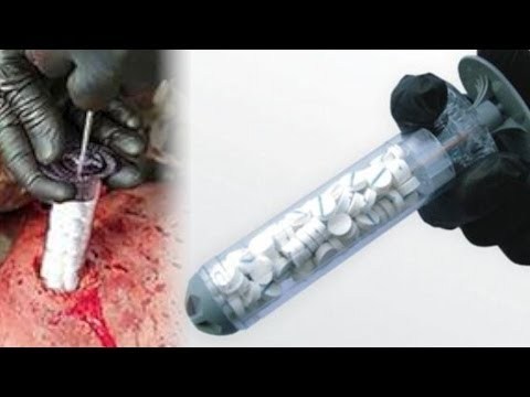
This new investigational device has made quite a splash during the past week. Manufactured by an Oregon company, it is designed to control bleeding, and is for use by combat medics and first responders.

Inspired by the old Fix-A-Flat expanding foam tire patch system, the XStat looks like a big syringe, and is filled with small 1cm sponges that expand rapidly when they get wet. It’s designed to stop hemorrhage in small wounds and wound tracts. Just pull back the plunger (which comes fully inserted to save space), push the unit into the wound, then hold the plunger while pulling the syringe out. This serves to leave the load of sponges in the tract and achieve rapid hemostasis.

image

It would seem that leaving a lot of tiny sponges in a wound could cause problems, especially if they are not removed at the time of definitive surgical management. However, each one is tagged with a radiopaque marker so they can be identified with xray or fluoro.

Preclinical trials have claimed to be successful, and an application has been submitted to the FDA for human use. This has the potential to save lives when bleeding gunshot wounds are encountered, especially in combat situations.

I have no financial interest in RevMedx, the manufacturer of this device.

More info: 

New Technology: The Next Generation Antibiotic Bead?

A number of surgical disciplines use antibiotic beads to deliver antimicrobial drugs to sites that may not have ideal serum penetration. Unfortunately, beads require multiple operations for placement and replacement until the desired effect is achieved.

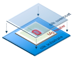
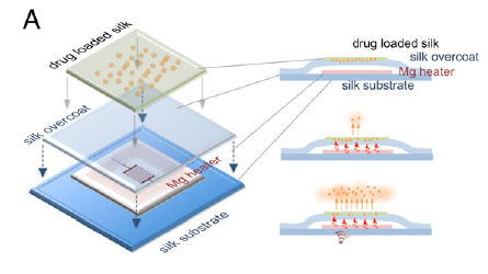
What if there was a way of delivering antimicrobial therapy directly to the tissues that works for up to two weeks, then dissolves with no trace? A system that does this is being developed by engineers at Tufts University and the University of Illinois at Urbana. They created a small magnesium coil that can be heated using magnetic induction. It is enclosed in a silk pocket and then implanted into the infected tissues. 

The tissues surrounding the device can be heated to different temperatures by placing an induction coil over it and delivering a specific amount of power.

It is also possible to deliver antibiotic doses directly to the tissue by embedding the drug into the silk pocket. As the coil heats up, the antibiotic is released from the fabric. 

The magnesium coil normally dissolves within a few hours when immersed in water, and it takes a bit longer when in direct contact with living tissue. The silk pocket prolongs the time to dissolution, depending on how thick it is. In the rat experiment described in the paper, there was little or no trace after 15 days.

Bottom line: This exciting technology has the potential to simplify the delivery of antimicrobial therapy directly to deeper tissues for extended periods, without the need for a second procedure to retrieve the device. We’ll see how this implant works in studies in larger animals. I’m sure other derivative applications are soon to follow.

Reference: Silk-based resorbable electronic devices for remotely controlled therapy and in vivo infection abatement. Proceedings in the National Academy of Sciences. Published online November 24, 2014.

What Percent Pneumothorax Is It?

What percent pneumothorax?

Frequently, radiologists and trauma professionals are coerced into describing the size of a pneumothorax seen on chest xray in percentage terms. They may say something like “the patient has a 30% pneumothorax.”

The truth is that one cannot estimate a 3D volume based on a 2D study like a conventional chest xray. Everyone has seen the patient who has no or a minimal pneumothorax on a supine chest xray, only to discover one of significant size with CT scan.

Very few centers have or use the software that can determine the percentage of chest volume taken up with air. There are only two percentages that can be determined by viewing a regular chest xray: 0% and 100%. Obviously, 0% means no visible pneumothorax, and 100% means complete collapse. Even 100% doesn’t really look like 100% because the completely collapsed lung takes up some space. See the xray at the top for a 100% pneumothorax.

If you line up 10 trauma professionals and show them a chest xray with a pneumothorax, you will get 10 different estimates of their size. And there aren’t any guidelines as to what size demands chest tube insertion and what size can be watched.

Bottom line: The solution is to be as quantitative as possible. Describe the pneumothorax in terms of the maximum distance the edge of the lung is from the inside of the chest wall, and which intercostal space the pneumothorax extends to. So instead of saying “the patient has a 25% pneumo,” say “the pneumothorax is 1 cm wide and extends from the apex to the fifth intercostal space on an upright film.”

Mystery Diagnosis: The Answer

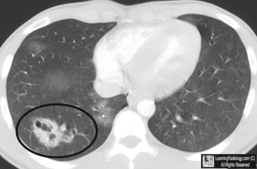
A young male suffered blunt torso trauma when struck by a car. Many of you sent your guesses for what is shown in the image below:

This patient sustained a traumatic pneumatocele. It is an uncommon injury in blunt trauma, and can also be caused by penetrating injury. It’s essentially a complicated laceration that fills with air leaking from torn airways (alveoli or bronchi of various sizes). 

There is usually some focal hemorrhage around the injury, which looks (and is) a pulmonary contusion. The hallmark is the bubble (or bubbles) of air that form in the area of the injury. Frequently, these can be seen on chest xray as well, although CT is much more sensitive. They are more commonly located near the pleural surface of the lung in blunt trauma, because this is the area of maximum impact. When present, they are often situated very close to a rib fracture.

Generally, these injuries do not require any specific management. They slowly heal over time, but it may take months for them to completely resolve.

Related posts: