AAST 2013: Screening For Blunt Cerebrovascular Injury

Blunt cerebrovascular injury (BCVI) is one of those low incidence, high consequence injuries like traumatic aortic rupture. Unfortunately, BCVI has gotten less attention and is less frequently screened for during trauma evaluation. There are two choices of screening criteria: digital subtraction angiography (DSA) or CT angiography (CTA). Previous work by the UT Memphis team showed that the sensitivity of evaluation by 32-slice CT angio is only 51%.

Although DSA has been considered the gold standard, there are several drawbacks. First, it is invasive and has its own collection of complications, including stroke. And it is not available at all centers, and even in those that have it, it is frequently a limited resource. Current 32-slice CTA risks missing significant injuries, which can also lead to stroke.

The UT Memphis team has now followed up their 32-slice work with a new study evaluating 64 slice scanners. Here are the factoids:

  • Over a one year period, 594 patients underwent both CTA and DSA for evaluation of BCVI
  • 128 patients had 163 injured vessels (!), 61% carotid and 39% vertebral
  • 5 DSA complications occurred (1%): 3 puncture site hematomas and 2 iatrogenic dissections
  • 64-slice CTA had an improved sensitivity of 68%

Bottom line: CT angiography for blunt cerebrovascular injury is following the same trajectory that it did for evaluating traumatic aortic injury. As the resolution of the scans improves, our diagnostic accuracy does the same. CTA using a 64-slice scanner will now begin to displace digital subtraction angiography due to its readier availability and low complication rate. 

BUT, the most important part of the process is that the trauma professional needs to use solid criteria to make the decision to use it!

Related posts:

Reference: Blunt cerebrovascular injury screening with 64-channel multidetector computed tomography: more slices finally cut it. AAST 2013, Paper 13.

AAST 2013: Nonselective Angiography For Splenic Injury

Angiography is yet another tool for use in the management of splenic injury. Over the past decade, it has helped improved splenic salvage rates into the mid-90% range. Angio can be used selectively (for very specific indications) or nonselectively (routinely).

The University of Tennesse – Memphis tried to examine whether there was any benefit to more frequent, nonselective use of this tool. They analyzed data in the National Trauma Data Bank (NTDB). They excluded patients with early splenectomy (within 6 hours). Selective vs nonselective use was determined by looking at the percentage of patients from a specific hospital that underwent the procedure. Low or no use hospitals used it from 0-20% of the time, and high use hospitals used it in >20% of cases. 

Here are the factoids:

  • 7412 records were analyzed
  • There was no difference in splenic salvage rates in low vs high angio use centers
  • Higher overall injury severity and AIS 5 for spleen was associated with higher delayed splenectomy

Bottom line: Not a very enlightening analysis. The researchers equated high utilization with non-selective utilization, which is a big flaw. The numbers are large, but the underlying assumptions are weak. And we already knew that higher injury severity and higher grade spleen injury equals higher splenectomy rate.

I recommend that any center that uses angiography as an adjunct to management of splenic injury to have specific indications (grade, degree of hemoperitoneum, CT contrast extravasation, etc). This ensures uniform use of this tool, and helps optimize the risk:benefit ratio to improve patient safety.

Related posts:

Reference: Are we lemmings?: Nonselective use of angiography provides no benefit in high-grade splenic injury. AAST 2013, Paper 12.

AAST 2013: Seizure Prophylaxis After TBI

The Brain Trauma Foundation has an extensive set of guidelines for the management of traumatic brain injury. One of these involves the routine administration of anti-seizure prophylaxis. The risk of seizure after TBI is higher than in the general population, and a high risk group has been identified (GCS<10, depressed skull fracture, cortical contusion, subdural / epidural / intracerebral hematoma, penetrating injury, or seizure within 24 hours.

This guideline showed that there was no data to support a recommendation with the highest degree of evidence, but it did suggest that anti-seizure meds decreased the incidence of early seizures (within 7 days), but these seizures had no effect on outcome. This meant that you might consider giving phenytoin, but there was always the risk associated with giving any new medication, and no real benefit to the outcome. This is one reason why the guideline is applied inconsistently by trauma professionals.

Researchers at the University of Florida – Jacksonville will be presenting a paper at AAST 2013 further questioning the advisability of this guideline. They retrospectively reviewed 93 patients who were admitted to their hospital for at least 7 days with severe blunt TBI. They split the patients into two groups, a control group (43 patients) who did not receive phenytoin and a study group (50 patients) who had therapeutic levels.

Here are the factoids:

  • More seizures occurred in the phenytoin group (4% vs 0%)
  • Mortality was the same (8% vs 7%)
  • Discharge disposition (home vs rehab) was the same
  • Hospital length of stay was longer in the prophylaxis patients (36 vs 25 days)
  • Glasgow Outcome Scale was worse in the phenytoin group (2.9 vs 3.4)

Bottom line: Don’t pay much attention to this study. It’s very small, making the quality of the results pretty shaky. There is no comment on matching other injuries that might account for a difference in length of stay. And is this the proper way to find a difference in Glasgow Outcome Scale, an integer value? What does 2.9 or 3.4 mean? Nothing. It has to be a 3 or a 4, no decimal point.

Although the data supporting the original Brain Trauma Foundation guideline is a bit weak, this paper does not support changing current practice. Needs more work (and more numbers).

Related post:

Reference: More Harm Than Good: Anti-seizure prophylaxis after traumatic brain injury does not decrease seizure rates but may inhibit functional recovery. AAST 2013 Paper 10.

Subdural Hematomas and Hygromas Simplified

There’s a lot of confusion about subdural pathology after head trauma. All subdural collections are located under the dura, on the surface of the brain. In some way they involve or can involve the bridging veins, which are somewhat fragile and get more so with age.

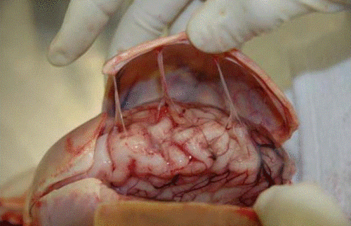
Head trauma causes a subdural hematoma by tearing some of these bridging veins. Notice how thick the dura is and how delicate the bridging veins are in the image below.

image

When these veins tear, bleeding ensues which layers out over the surface of the brain in that area. If the bleeding does not stop, pressure builds and begins compressing and shifting the brain. A subdural hematoma is considered acute from time of injury until about 3 days later. During this time, it appears more dense than brain tissue.

After about 3-7 days, the clot begins to liquefy and becomes less dense on CT. Many hematomas are reabsorbed, but occasionally there is repeated bleeding from the bridging veins, or the hematoma draws fluid into itself due to the concentration gradient. It can enlarge and begin to cause new symptoms. During this period it is considered subacute.

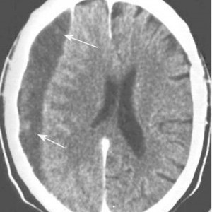
It moves on to a more chronic stage over the ensuing weeks. The blood cells in it break down completely, and the fluid that is left is generally less dense than the brain underneath it. The image below shows a chronic subdural (arrows).

image

Hygromas are different, in that they are a collection of CSF and not blood. They are caused by a tear in the meninges and allow CSF to accumulate in the subdural space. This can be caused by head trauma as well, and is generally very slow to form. They can lead to slow neurologic deterioration, and are often found on head CT in patients with a history of falls, sometimes in the distant past. CT appearance is similar to a chronic subdural, but the density is the same as CSF, so it should have the same appearance as the fluid in the ventricle on CT.

Related posts:

AAST 2013: Practice Guideline For Open Fractures

Evidence based guidelines continue to be part of the practice of medicine. They seek to standardize what we trauma professionals do to manage common clinical problems. A new orthopedic guideline was evaluated at the University of Michigan and the results are being reported at the upcoming AAST meeting in San Francisco.

There is considerable variation in the management of open fractures, ranging from timing of washout/repair, grading, and antibiotic management. The U of M group standardized the way they administered antibiotics in these patients. They implemented a protocol as follows:

  • Gustilo Grade I or II – cefazolin for 48 hours (clindamycin if allergic)
  • Gustilo Grade III – ceftriaxone for 48 hours (clinda or aztreonam if allergic)
  • No aminogyclosides, penicillin or vancomycin

They studied their results in 174 patients with open femur or tib/fib fractures (101 pre-protocol and 73 post; one apparently had both areas fractured). Risk adjusted surgical infection rates were calculated using the National Health Safety Index risk index, which is calculated using the ASA score, the wound classification, and the duration of the operative procedure. 

Here are the factoids:

  • The use of aminoglycosides and vancomycin decreased from 54% to 16%
  • Skin and soft tissue infection rates were not different (21% pre and 25% post)
  • People did not change their fracture grading to “game” the system
  • Infections with antibiotic resistant organisms or MRSA were similar in the two groups
  • The authors did not report time to operation in these open fractures

Bottom line: This is a good first shot at standardizing antibiotic use in patients with open fractures. The numbers are very small, and time to OR was not taken into account. Whereas the 8 hour rule for open fractures was dogma and has pretty much been discounted, antibiotic use is a case of “every man for himself.” It is important to continue this work, because I’m sure there will be cost and education benefits from following a protocol like this. More numbers need to be generated, and anyone who adopts this protocol now needs to closely watch their soft tissue and bone infection results in their PI process.

Related posts:

Reference: Evidence-based protocol for prophylactic antibiotics in open fractures: improved antibiotic stewardship with no increase in infection rates. AAST 2013 Paper 62.