New Trauma MedEd Newsletter Released Tonight To Subscribers!

The September issue of Trauma MedEd is ready! Subscribers will receive it tonight. This issue is devoted to prevention

Included are articles on:

  • Motorcycle helmets
  • Elderly falls
  • Dug use
  • Prevention map mashups
  • And more!

As mentioned above, subscribers will get the issue delivered tonight to their preferred email address. It will be available to everybody else later this week on the blog.

Check out back issues, and subscribe now! Get it first by clicking here!

Is Repeat Head CT In Pediatric Trauma Really Necessary?

During the past several years, it has finally begun to dawn on us that radiation is not very good for us. This is especially true in pediatric patients, where they may not feel the full effect of over-irradiation for years to come. We’ve made efforts to decrease or eliminate studies when possible. We’ve adopted the principles of ALARA (doses as low as reasonably achievable).

But there are some areas of the body that demand evaluation using CT after blunt trauma. The head is one, since MRI is not as immediately available, studies take a long time, and they may demand sedation to the point of intubation in smaller children.

Unfortunately, we have found another way to increase radiation exposure when using head CT. The repeat scan. The concern is that pathology noted on the first scan may worsen, indicating that a more specific (and usually invasive) intervention is needed. 

A study was presented at the AAST this year examining the use of specific clinical criteria to determine whether a repeat head CT was necessary after blunt head trauma in children. Here are the factoids:

  • This was a retrospective cohort review of a pool of 435 patients admitted to a pediatric trauma center over 8 years (!) with accidental TBI
  • Only 120 were eligible (!), with both some type of intracranial hemorrhage and a GCS of 14-15
  • Fourteen patients did not receive repeat head CT; the remaining 106 did
  • Pediatric age was not defined in the abstract, but repeat CT kids were older than no repeat kids (8 years vs 3 years)
  • None of the children not receiving a repeat scan had worsening symptoms
  • Seven children with repeat CT worsened; 5 had an epidural and 2 had subarachnoid hemorrhage. One of the epidurals showed an increase in mental status

Bottom line: Another poorly constructed study. Although it agrees with my bias toward using physical exam in place of repeat CT in select adult head trauma, I just can’t add this study to my personal library. There is a small but growing body of literature about this in adults, but this pediatric one is nowhere good enough to alter my practice. Too few patients, retrospective (we don’t know why they chose to scan or not scan), and major age variances are only a few of the problems. And the fact that one child with a repeat scan and no change in exam showed an increasing epidural that needed operative intervention is frightening.

Let’s keep looking for ways to decrease radiation exposure. But first, let’s craft some really good, prospective studies so we can be sure we’re not jeopardizing the well-being of our children.

Related posts:

Reference: Is routine repeat brain CT necessary in all children with mild traumatic brain injury? AAST 2013, paper 56.

When To Image The Aorta In Blunt Trauma

Blunt injury to the thoracic aorta is one of those potentially devastating ones that you (and your patient) can’t afford to miss. Quite a bit has been written about the findings and mechanisms. But how do you put it all together and decide when to order a screening CT?

There are a number of high risk findings associated with blunt aortic injury. Recognize that they are associated with the injury, but are still not very common. They are:

  • Fractures of the sternum or first rib
  • Wide mediastinum
  • Displacements of mediastinal structures (left mainstem down, trachea right, esophagus right)
  • Loss of the aortopulmonary window
  • Apical cap over the left lung

Here’s a sensible method for screening for blunt aortic injury, using CT scan:

  • Reasonable mechanism (fall from greater than 20 feet, pedestrian struck, motorcycle crash, car crash at “highway speed”) PLUS any one of the high risk findings above.
  • Extreme mechanism alone (e.g. car crash with closing velocity at greater than highway speed, torso crush)

Note on torso crush: I have seen three aortic injuries from torso crush in my career, one from a load of plywood falling onto the patient’s chest, one from dirt crushing someone when the trench they were digging collapsed, and one whose chest was run over by a car.

What Is A Wide Mediastinum Anyway?

Trauma professionals are always on the lookout for injuries that can kill you. Thoracic aortic injury from blunt trauma is one of those injuries. Thankfully, it is uncommon, but it can certainly be deadly.

One of the screening tests used to detect aortic injury is the old-fashioned chest xray. This test is said to be about 50% sensitive, with a negative predictive value of about 80%. However, the sensitivity is probably decreasing and the negative predictive value increasing due to the rapidly increasing number of obese patients that we see.

A wide mediastinum is defined as being > 8cm in width. In this day and age of digital imaging, you will need to use the measurement tool on your workstation to figure this out.

Unfortunately, it seems like most chest xrays show wide mediastinum these days. What are the most common causes for this?

  • Technique. The standard xray technique used to reduce magnification of the anterior mediastinum (where the aortic arch lives) is a tube distance of 72 inches from the patient, shot back to front. We can’t do this for trauma patients because we can’t stand them up and are reluctant to prone them. The standard trauma room technique is 36 inches from the patient shot front to back. This serves to magnify the mediastinal image and make it look wide.
  • Obesity. The more fat in the mediastinum, the wider it looks. The more fat on the back, the further the mediastinum is from the xray plate and the greater the magnification.
  • Other mediastinal blood. Major blunt trauma to the chest can cause bleeding from small veins in the mediastinum, making it look wide.
  • Thymus. Only in kids, though.
  • Aortic injury. Last but not least. Only a few percent of people with wide mediastinum will actually have the injury.

If you encounter a wide mediastinum on chest xray in a patient with a significant mechanism for aortic injury, then they should be screened using helical CT.

Related post:

What The Heck? Final Answer!

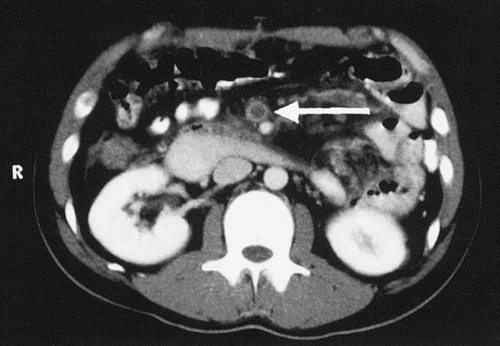
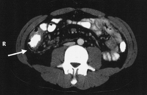
This case involved a young male who had undergone a splenectomy for blunt trauma. He had an unremarkable postop course and was discharged after 5 days. He presented 2 weeks later with abdominal pain out of proportion to his exam. 

The first CT scan hint showed pneumatosis of the cecum, which is decidedly unusual in a young person:

The next CT scan hint showed thrombosis in the superior mesenteric vein:

This case illustrates an uncommon yet dangerous complication of splenectomy: portal and mesenteric veinous thrombosis. It is generally believed to be caused by thrombocytosis resulting from the splenectomy. The pneumatosis is most likely due to problems resulting from venous congestion.

In this particular case, the patient’s abdominal exam was not consistent with a catastrophic problem (yet). He was taken quickly to the interventional radiography department, where a catheter was inserted and lytic therapy was begun. HIs pain resolved, and repeat CT showed recanalization of much of his portal and mesenteric venous system, as well as resolution of the pneumatosis. He was discharged on warfarin therapy.

Bottom line: Uncommon and dangerous: a bad combination. This often results in delayed diagnosis and severe abdominal complications, including loss of much of the intestine. Monitor the platelet count closely postop until it plateaus, and if it still rising on discharge from the hospital, arrange for monitoring as an outpatient. Dogma tells us to treat with aspirin or similar drugs as the count approaches 1 million/cubic mm, but good studies are lacking. Development of this particular complication requires anticoagulation, and possibly lytics or surgical resection depending on patient condition.


Hat tip to StillChucklesandNP for getting the answer with limited information, and whom I had the pleasure of meeting on a recent visit to her hospital!