Arab Health 2013 – Dubai

Heads up to all my readers in the Middle East! I’m attending the Arab Health healthcare exhibition and congress in Dubai at the end of January.

This huge event is in its 38th year, making it the longest running meeting in the area. It will be located at the Dubai International Convention and Exhibition Centre, from Jan 28-31. I’ll be at the Minnesota International Medicine exhibit located in Hall 1 on Monday through Wednesday. Stop by and see me! I’d love an opportunity to meet my readers! I would also enjoy an opportunity to visit the Trauma Centre at Rashid Hospital, if possible!

Feel free to email, tweet or Skype me (skype:Michael.D.McGonigal)

Next TraumaMedEd Newsletter Available Soon!

Well, the holidays slowed me down a bit, but the December issue of TraumaMedEd is finally ready to go! It’s another How To issue, with tips on stabs, occult pneumothorax and more!

Subscribers will get the issue delivered tomorrow evening (Central time). It will be available to everybody on the Thursday blog post.

Check out back issues, and subscribe now and get it first by clicking here!

Trauma Mythbusters: Spinal Cord Injury From Airway Management

Airway management is one of the most anxiety provoking procedures performed by trauma professionals. The main fear is that the airway will be lost during attempts to secure it and patient demise will soon follow. Add some facial fractures, bleeding, and an “unfavorable body habitus”, and the average prehospital or ED provider is really on edge.

The next most common fear is that providing a definitive airway in a patient with a known (or even suspected) cervical spine injury could cause a catastrophic neurologic injury. This was first addressed back in 1989 (before the time of video laryngoscopy, and when flexible scopes were rarely available in the ED). The authors found no verifiable reports of such an injury in the entire English literature

Over the years, a few case reports have cropped up. As in so much of the medical literature, causality is hard to prove. The patient was normal before anesthesia, and afterwards they were not. Had to be the intubation, right?

Not so fast! Let’s break it down and look at what we do know. Biomechanical studies have shown that the manipulation that occurs in direct laryngoscopy isn’t as bad as it looks. Studies in uninjured models are enlightening (minimal movement with blade insertion, slight rotational movements with blade elevation, and a little more rotation during the intubation). Most of this (slight) movement occurs from occiput to C2, with little motion at all at C3 and below.

But that was on an uninjured model. What about ones that simulate an injured spine? Specifically an injury in the upper spine area that we know moves?

  • Cricoid pressure caused no appreciable changes in the spine
  • Chin lift and jaw thrust reduced space available for the cord (SAC) by 1 and 2.5mm, respectively, and caused an angulation of about 4-5 degrees
  • SAC narrowed by only 1.5mm, even with maximum flexion and extension
  • Oral and nasal intubation narrowed SAC by 1.6mm, and resulted in a maximum of 2.5 degrees of rotation
  • Video laryngoscopy results in about half of the rotational movement of direct laryngoscopy
But what about these sporadic reports of neurologic deficits after intubation? What is often neglected is that spinal blood flow and long-term neck positioning have a major impact on cord function. Even relatively mild malpositioning of the cervical spine for extended periods during an OR case have been documented. 

Bottom line: From a mechanical standpoint, even in unstable spine models, the maneuvers we use in preparation for intubation cause more movement of the spine than does the intubation procedure itself. The true number of spinal cord injuries actually (and provably) caused by intubation approaches zero. The literature suggests that video laryngoscopy results in less overall movement during intubation, but it doesn’t seem to have an impact on cord injury (you can’t get less than zero). 

References:

  • Spinal cord injury and direct laryngoscopy – the legend lives on. Br J Anesth 84(6):705-709.
  • Airway management in adults after cervical spine trauma. Anesthesiology 104(6):1293-1318, 2006.

Best Of: Why Did The Trauma Team Cut Off My Clothes?

The fifth highest priority taught in the ATLS course is exposure. This generally means getting the patient’s clothes off so any hidden injuries can be identified. Early in my career, I was called to see a patient who had a gunshot to the chest that had been missed because the consulting physician had neglected to cut off her bra. A small caliber wound was found under the elastic strap in her left anterior axillary line after a chest xray showed a bullet in mid-thorax.

The usual trauma activation routine is to cut off the clothes. There are several tips and tricks we use to do this quickly. And a number of commercial products are out there to make it even easier.

But do we really need to cut everyone’s clothes off? I’m not disputing the fact that it’s important to be able to examine every square inch. But do we need to destroy everything our patient is wearing? I once saw a sequined wedding dress cut off (it’s almost as bad as cutting off a down jacket).

The answer is no. The key concept here is patient safety. Can you safely remove the clothing in a less destructive way? For most victims of major blunt trauma, we worry a lot about the spine. Unfortunately, it’s just not possible to allow the patient to wriggle out of their clothes and protect their spine. The same goes for fractures; it may be too uncomfortable to remove clothing because of fracture movement so scissors are required.

Penetrating trauma is a bit different, and in many cases it’s a good idea to try to get the clothing off intact. Once again, if spinal injury is a consideration (gunshots only), the involved clothes should be cut off. A patient with a gunshot to the chest can probably have their pants safely and gently pulled off, but their shirt and coat must be cut.

The police forensic investigators like to have intact clothing, if possible. This is another good reason to try to remove clothing from penetrating injury victims without cutting. 

Bottom line: Think before you cut clothes! Major blunt trauma and bad injuries require scissors. Lesser energy blunt injury may allow some pieces of clothing to be removed in the usual method. Most penetrating injury does not require cutting. But if you must (for patient safety), avoid any holes in the fabric so forensics experts can do their job.

What Is: Venous Sinus Thrombosis?

Venous sinus thrombosis is in the news now, since Hillary Clinton is currently hospitalized and being treated for it. Most trauma professionals have probably never heard of it, let alone seen one. Here’s a quick primer on what you need to know about this problem.

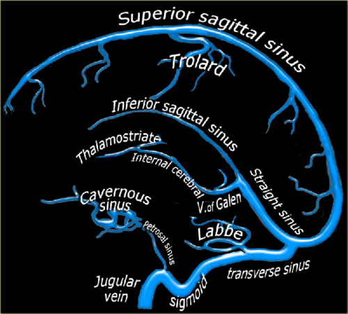
Blood circulation from the cerebral veins drains into the various venous sinus systems around the brain. Additionally, CSF is reabsorbed by the arachnoid villi, which in turn drain into the superior sagittal sinus. A clot in any part of this sinus system (see image below) results in a partial or complete obstruction, which can lead to rapidly increasing pressure in the brain.
image

The condition is very rare, with only about 3 cases per million people recognized. Although most occur in people with clotting abnormalities, trauma is always on the list. The most common traumatic issue is head injury, and almost always involves a skull fracture near the involved venous sinus.

Clinically, awake patients complain of a steadily increasing headache that is not touched by any of the usual analgesics. Some describe a headache with very sudden onset, the so-called “thunderclap headache.” Any number of neurologic abnormalities can then occur, ranging from nothing to seizures to rapid onset of coma.

Diagnosis is difficult. CT scan unreliably shows the lesion, and subtle venous changes may be overshadowed by findings from the original trauma. However, it should be performed first to accurately diagnose the more common problems that are most likely to be present. If the CT comes up clean, the gold standard is MRI with magnetic resonance venography (MRV)

Patients with significant swelling problems may require hyperosmotic therapy and/or decompressive craniectomy. Milder cases can be treated with anticoagulation, but a thorough risk/benefit assessment must be carried out. Remember, these people usually have brain injury that has resulted in bleeding somewhere it shouldn’t be, so anticoagulation can be dangerous! A few rare case reports of thrombolysis (!!) and clot extraction are out there, too, but these are for extreme cases only.

Bottom line: It’s likely that Ms. Clinton suffered a temporal bone fracture from her fall, and did well initially like most patients. However, increasing symptoms (headache), likely prompted a repeat CT followed by the MRI which showed thrombosis of her transverse sinus. Luckily, she did not have significantly increased intracranial pressure leading to more serious neurologic problems. Judicious anticoagulation with warfarin and very close monitoring were instituted, and seem to be working well so far.

References:

  • Thrombosis of the cerebral veins and sinuses. NEJM 352:1791-1798, 2005.
  • Cerebral venous sinus thrombosis complicating traumatic head injury. J Clin Neurosci 19(7):1058-1059, 2012.