Angiography And Splenic Salvage

Variations in the way we deal with trauma can have a significant impact on patient outcome. This has been documented most recently in the use of angioembolization when dealing with patients with spleen injuries. The first paper presented at EAST 2013 looked at outcomes at hospitals that use angio more heavily vs those who don’t.

They analyzed 1275 patients presenting to 4 Level I trauma centers. Two centers were high-use (11% and 19% usage) and the other 2 were low-use (1% and 4%). The outcomes studied were the splenic salvage rate and success in nonoperative management. And although patients at the low angio use centers had a higher ISS, the splenic injury grade was the same.

Interesting findings included:

  • Admission splenectomy rate was the same, meaning that both types of centers used the same criteria when the patient rolled through the door
  • High angio use centers had higher overall salvage rates (82% vs 77%)  and greater success with nonoperative managment (96% vs 92%)
  • In high grade injury (grade 3 and 4) the salvage rate was still better (67% vs 56%) and nonop success rates were much better (92% vs 80%)
  • In patients who were initially managed nonoperatively, use of angio was associated with salvage
  • Patients in high angio centers were more likely to leave the hospital with their spleen where it should be
  • There was no analysis of complications from angiography
  • There was no comment on how these patients were managed on a day to day basis

Bottom line: There is a considerable amount of variation in how trauma centers use angiography for spleen injury. Unfortunately, this variability is allowing people to lose their spleens at centers who don’t use it as much. The overall success rate in managing spleen injury (all comers) has historically been about 93%. More aggressive use of angiography is now shown to improve that to 97%. Given this new data, angio needs to be considered in patients with grade 3+ injury and in any with contrast extravasation. And the overall management should be standardized as well.

Reference: Variation in splenic artery embolization and spleen salvage: a multicenter analysis. Paper 1, EAST annual scientific assembly, Jan 15, 2013.

EAST Starts Tomorrow!

I’m off to Phoenix for the Eastern Association for the Surgery of Trauma (EAST) annual scientific assembly. I’ll be tweeting continuously during the various scientific sessions using the hashtag #east2013. And I’ll select the most interesting presentations daily and post more in-depth discussions of them here. I’ll review the new and revised trauma practice guidelines next week, and I’ll probably have a few more papers to discuss over the coming weeks.

For any fans attending the meeting, please stop me and say hi!

The Referral Hospital Trauma Rule

The majority of trauma patients are seen initially at non-trauma centers. And the majority of those patients can be treated just fine at that local hospital. However, a few (some say about 15%) do need to be transferred. The question frequently arises, “what studies do I need to do before transferring?

The danger is that doing things that slow down the transfer can result in bad outcomes. For example, a patient may have a spleen injury that is actively bleeding. Every minute that this patient is not receiving “definitive treatment”, she loses more blood. And every cc of blood lost causes her to inch closer to shock, other complications, or death.

The key is to get people who need a higher level of trauma care on their way to a higher level trauma center as soon as the need is recognized. There is a natural tendency to do diagnostic studies, such as CT scan, in these patients. Sometimes they are needed to actually figure out what is going on. But more often they are obtained to “do a complete workup” or because “the trauma center expects me to.”

Unfortunately, these are incorrect assumptions. The complete workup cannot be used by the referral center if they are shipping the patient, and for a variety of reasons they may not be useful to the trauma center. This is one of the major reasons that referral patients receive extra radiation exposure. About half of the studies performed at the referral hospitals need to be repeated!

The Referral Hospital Trauma Rule: Do any simple study needed to ensure the patient will stay alive until the helicopter/ambulance arrives (typically chest or pelvic xray). If at any point, you see something obviously not treatable at your hospital (i.e. open fracture, GCS 8, partial amputation), DO NO FURTHER STUDIES AND PREPARE TO TRANSFER. If the patient does not have such an obvious problem, do only the tests you need to determine if you can keep the patient. But as soon as you find anything that you cannot treat, stop further studies and prepare to send the patient onward. And don’t forget to send working copies of the few studies that you did get.

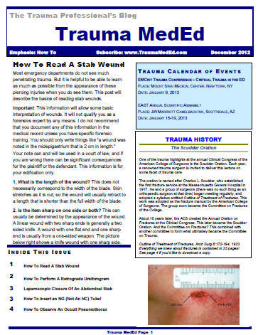
December TraumaMedEd Newsletter

The December newsletter is here! Click the image below or the link at the bottom to download. This month’s topic is How To, providing practical tips on things like:

  • “Reading” stab wounds
  • Performing a retrograde urethrogram better than a urologist
  • Closing abdominal stabs using a laparoscope
  • Properly inserting an NG tube
  • Managing occult pneumothorax

Subscribers had the newsletter emailed to them on Tuesday. If you want to subscribe (and download back issues), click here.

Download the newsletter

Compliance With Bladder Injury Guidelines

Management of bladder injury seems straightforward. For many years, the gold standard was the cystogram. But for best results, this study had to be done a certain way. This included bladder instillation, imaging in two planes (AP and lateral), and a post-void view.

As CT scan use became more prevalent, we discovered that diagnosis of bladder injury became even more accurate. However, we also soon discovered that even though the pictures looked very good, some bladder injuries were being missed. It turns out that allowing passive filling of the bladder by clamping the urinary catheter (or not inserting one) missed upwards of 50% of injuries.

So the standard became the “CT cysto” technique. It is performed similar to a non-CT cystogram, by infusing contrast into the bladder under pressure. With this modification, the accuracy of the study approaches 100%. But do we all do it this way? No!

A study from the University of Utah reviewed registry data from all Utah Level I trauma centers over a 15 year period. A total of 124 patients with bladder injuries were identified. Interesting results include:

  • Extraperitoneal rupture was more common (60%) than intraperitoneal (31%) or both (9%)
  • Conventional CT was used in 56%, and cysto or CT cysto in 24%. The remainder were found in OR.
  • Initial imaging missed or incorrectly diagnosed this injury in 13% of patients! Nine of these used the wrong study (conventional CT), but 4 of these missed occurred using the recommended one.
  • Overall compliance with using the recommended study was only 44%

Bottom line: Compliance in this 15 year study was low. Unfortunately, they lumped conventional cystogram with CT cysto. These days, fewer conventional studies are performed and the error rate may be higher. However, current day compliance is still low in my experience. A bladder evaluation guideline should be developed and disseminated to emergency physicians, surgeons and radiologists (see the CT cysto link below). This is the only way we’ll be able to decrease the number of missed injuries for this problem.

Related posts:

Reference: Process improvement in trauma: traumatic bladder injuries and compliance with recommended imaging. J Trauma 74(1):264-269, 2013.