Pelvic Fractures: OR vs Angio In The Unstable Patient

One of the cardinal rules of trauma care is that hemodynamically unstable patients can only go the the operating room from the ED. No trips to CT, xray, etc. Trauma professionals occasionally try to make exceptions to the rule, but it usually doesn’t work out.

Well, what about the patient with severe pelvic fractures who is or becomes unstable? Pelvic fracture bleeding is not always easy or even possible to control in the OR, and angiography offers a way to identify and stop the bleeding, right?

The trauma group at Ryder in Miami did a lengthy (13 year) retrospective review of their experience with these patients. They looked at every patient who underwent angiography, then identified the subset that went to the OR followed by angiography. There were 134 angio patients and 49 OR to angio patients on whom they based their analysis. Obviously, there is plenty of opportunity for bias in this study, and many of the study patients identified had to be excluded due to incomplete records.

Patients who went to the OR first tended to have similar injury severity but were sicker than the angio alone group. Crystalloid and blood resuscitation volumes were significantly higher in the OR group as well. Most of these patients underwent a laparotomy, and 64% had active intra-abdominal bleeding. None died in OR, and most were left with a damage control abdominal closure.

In the angio group, there were really 2 subsets: angio alone, and angio followed by OR. Mortality in the angio alone group was similar to the OR-angio group. But deaths skyrocketed in those who went from angio to OR (67% vs 20%). This is likely due to them failing angiographic management of bleeding. Three patients died in the angio suite.

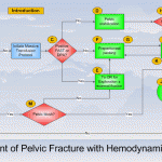
Bottom line: There’s a lot of data in this paper, and some of the results can be explained by selection bias. However, they appear to support algorithms released by EAST and the WTA (see diagram above). In general, a trauma patient with severe pelvic fractures and hemodynamic instability needs to go to OR to identify and treat any source of intra-abdominal bleeding. If pelvic bleeding remains a problem, preperitoneal packing may be considered, followed by a trip to angio at that point. The rule that unstable patients should only go to OR (or an ambulance bound for a trauma center if there is no OR) still holds!

Reference: Operating room or angiography suite for hemodynamically unstable pelvic fractures? J Trauma 72(2):364-372, 2012.

Quiz: There is just one extremely rare reason that I know of to move to CT with a hemodynamically unstable trauma patient. Leave a comment with your guess.

Central Line Insertion Causes Hypercoagulability?

Again, I’m not a fan of animal studies. But this one, presented at EAST 2012, involves both pigs and humans and is so intriguing I just have to share it. The authors have a track record of studying coagulation issues with thromboelastography (TEG) in both animals and people. They previously showed that hypercoagulability detectable by TEG occurs after insertion of pulmonary artery catheters in swine and critically ill humans.

In this follow-on study, they looked at TEG profiles in 16 healthy swine and 8 critically ill humans after insertion of a central venous catheter (CVC). They found that CVC insertion induced the same type of hypercoagulable state. TEG clotting time and initial clot formation time decreased, and fibrin cross-linking accelerated. The changes were somewhat less in humans, but were still significant in both groups. All coag tests (PT, PTT, INR) and measured coag factors (von Willebrand, AT III) were unchanged.

Interestingly, in the animal group the hypercoagulable state persisted for at least 3 hours after CVC removal. And the hypercoagulability could be prevented with enoxaparin, but not heparin.

Bottom line: The idea that hypercoagulability could be induced by central arterial or venous catheter placement is intriguing, although this work has not been replicated by others yet. What if hypercoagulability occurs with any invasion of the vascular system? We may eventually discover that the increased incidence of DVT we have been fighting in the hospital setting is in part due to our ubiquitous use of IVs and routine blood draws.

Reference: Insertion of central venous catheters induces a hypercoagulable state. Presented at the 25th Annual Scientific Assembly of EAST, Orlando FL, 2012.

By Request: Tranexamic Acid In Trauma

I have received several requests to write about tranexamic acid (TXA) and trauma patients. There continues to be a lot of interest in this agent, especially in the military, and there are some good, recent trauma papers to review. Additional papers are being published on its use for control of bleeding, mostly in non-trauma journals.

Tranexamic acid works differently than the quick clotting agents out there. It is anantifibrinolytic, so it actually prevents clot breakdown. It has been approved by the FDA for use in hemophiliacs undergoing dental work and for menorrhagia. Thrombotic complications have been described, so it cannot be used with prothrombin complex concentrate or recombinant activated factor VII.

The most recent and best known study on TXA is the CRASH-2 study. It was extremely well designed and included over 20,000 patients in hospitals spanning 40 countries. The study design has survived serious scrutiny. They found that TXA use in trauma patients reduced the relative risk of death by 9% (from 16% to 14.5%). The risk of death specifically from bleeding was reduced by 15%. And use of TXA in the most severely injured patients, those who would die of bleeding on the day of randomization, was reduced by 20%. There were no adverse events or differences in thrombotic events, including deep venous thrombosis.

Bottom line: TXA has been shown to be effective, safe and inexpensive (about $200 for treatment using retail pricing). It is the only drug that has been shown to reduce all-cause and mortality from bleeding in a high quality trial. It has already been adopted by some hospitals in both the US and the UK. CRASH-2 suggested that TXA was of most benefit when given within 3 hours of injury and in patients with a systolic pressure less than or equal to 75 torr. Trauma centers should begin incorporating this important drug into their initial treatment protocols now.

Final thought: Unless new studies uncover major flaws with this drug, it will eventually be started by EMS in the field in select cases.

Reference: Tranexamic acid for trauma patients: a critical review of the literature. J Trauma 71(1):S9-S14, 2011.

A Better Way To Repair Nerve Injuries?

I’m usually not too keen on animal studies. Many times, the translation from animal to human just doesn’t work out. However, a study published last week on nerve injury caught my interest.

Nerves don’t heal well. The problem is that the neurons at the injured nerve endings tend to die back. Small vesicles form that seal the damaged ends, such that repairs that occur after sealing do not reliably re-establish continuity.

A group of researchers at the University of Texas at Austin and Vanderbilt experimented with manipulating the micro-environment at the injured ends of the nerve to control vesicle formation and action. 

The researchers have practiced a new technique on about 200 rats, severing a number of peripheral nerves, including the sciatic. After injury, a calcium-free hypotonic salt solution containing methylene blue (an antioxidant) was injected into the area. This shuts down the mechanism for vesicle formation, preventing the body from prematurely closing the cut ends of the neurons.

Next, the cut nerve ends were approximated using microsuture technique without any sharp debridement. Polyethylene glycol was then injected into the repaired area. This serves to dehydrate the outer membranes of the neurons and to induce them to flow back together. Finally, an isotonic calcium rich solution was applied to the repair to induce vesicle formation and fusion to close any remaining holes in the plasmalemma.

The video above shows the remarkable results. Most rats began moving the injured extremity immediately upon awakening from anesthesia. Nearly all had regained 60-70% of function within 2-4 weeks of injury.

Bottom line: If this pans out (in the somewhat distant future), expect to see possible interventions beginning in the field immediately after injury, starting with injections of the hypotonic calcium-free solution. Surgeons will take it from there. This is an animal study to keep an eye on!

Video: Rats walking after sciatic nerve injury (courtesy NewScientist.com)

Reference: Rapid, effective, and long-lasting behavioral recovery produced by microsutures, methylene blue, and polyethylene glycol after completely cutting rat sciatic nerves. J Neuroscience Res, published online 3 Feb 2012.