The Future of the Medical Journal

Journals have been printed on trees since forever. Within the past 10 years, there has been a shift toward also making this content available on the publishers’ web sites. Now, Lippincott Williams & Wilkins (LWW), publisher of the journal Neurosurgery (and many others) has taken an important step forward.

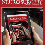
Beginning with the current issue (March 2012), this journal is now available within an iPad app. One of the problems with the traditional journal was that you had to go to the library to read it. And medical libraries can’t carry every journal, so if the one of interest was not in the collection, you had to wait a few days for the librarian to get a copy.

With the onset of the internet age, journals began to appear online. Most major publishers had a web presence and made their journals available online. However, most provided full text content for free only to their subscribers. At best, a non-subscriber would get the abstract, or an occasional free article. Biomedical libraries typically subscribed to services where hundreds of journals were available as full text to medical staff members, residents and students. The downside was, and still is, that you need access to a desktop to comfortably consume the articles. The web sites were just not that friendly for the small screen.

Finally, LWW has made a concerted effort to provide this content in tablet format. More and more trauma professionals are carrying these devices and using them in their practices. This brings the content closer to the patient and provides it in a very consumable format. It also allows the publisher to add dynamic digital content (audio and video) to the material.

I’ve been using this app for about a day and am impressed. Here are some key features:

  • Portability is excellent. A continuous internet connection is not needed. The entire journal issue is downloaded and added to your library, and you are only limited by the amount of storage on your iPad.
  • Extra digital content is routine. And since it is already downloaded with the issue, there is no waiting for it to load.
  • Articles have the same look and feel as the journal and can be panned or zoomed for readability.
  • Content sharing is possible (somewhat). Each article allows the user to share on Twitter or Facebook. The article can also be emailed to others. However, only a link to the article is provided, and if you are not a journal subscriber you’ll either have to pay up or use your medical library account to get full text when the link is opened.

What doesn’t it do? There is currently no ability to search through an article or issue. And it would be nice to interact with one of the authors via an email link to ask questions or make suggestions. Finally, Apple is not the only tablet maker out there. Publishers will need to make sure apps are available on the Android platform as well.

Bottom line: This is a great first-try at moving journals onto a mobile platform. I expect that LWW will begin to roll out this format for all their journals since they’ve now figured out how to do it. And expect all the other publishers to jump on the bandwagon as well. Journals are eventually going to go the way of DVD movies, and we’ll end up streaming our professional content from some company with a big red logo.

Reference: Search for “Neurosurgery” in the Apple App Store.

New Technology: Fracture Putty

Fracture healing takes a long time, as many of our patients can attest to. Six or eight weeks, and even more may be required for full healing. Researchers at the University of Georgia and in Houston have completed an animal study on rats using a type of “fracture putty” that dramatically speeds up this process. 

The researchers used adult mesenchymal stem cells that produce a protein which is involved in bone healing and regeneration. They created a gel using these cells, and injected them into the fracture sites which were stabilized externally (imagine a rat external fixator!). The fractures healed rapidly, and within 2 weeks the rats could run and stand on their legs normally.

Bottom line: The next step is to translate this work to larger animals. Strength and durability are major concerns. The amount of stress placed on rat legs and human legs is considerably different. If this pans out, it could revolutionize fracture healing, especially in cases where there may be highly disabling segmental bone loss (read: military). It will be several years before this can move to human studies.

Reference: University of Georgia Regenerative Bioscience Center

Why Did The Trauma Team Cut Off My Clothes?

The fifth highest priority taught in the ATLS course is exposure. This generally means getting the patient’s clothes off so any hidden injuries can be identified. Early in my career, I was called to see a patient who had a gunshot to the chest that had been missed because the consulting physician had neglected to cut off her bra. A small caliber wound was found under the elastic strap in her left anterior axillary line after a chest xray showed a bullet in mid-thorax.

The usual trauma activation routine is to cut off the clothes. There are several tips and tricks we use to do this quickly. And a number of commercial products are out there to make it even easier.

But do we really need to cut everyone’s clothes off? I’m not disputing the fact that it’s important to be able to examine every square inch. But do we need to destroy everything our patient is wearing? I once saw a sequined wedding dress cut off (it’s almost as bad as cutting off a down jacket).

The answer is no. The key concept here is patient safety. Can you safely remove the clothing in a less destructive way? For most victims of major blunt trauma, we worry a lot about the spine. Unfortunately, it’s just not possible to allow the patient to wriggle out of their clothes and protect their spine. The same goes for fractures; it may be too uncomfortable to remove clothing because of fracture movement so scissors are required.

Penetrating trauma is a bit different, and in many cases it’s a good idea to try to get the clothing off intact. Once again, if spinal injury is a consideration (gunshots only), the involved clothes should be cut off. A patient with a gunshot to the chest can probably have their pants safely and gently pulled off, but their shirt and coat must be cut.

The police forensic investigators like to have intact clothing, if possible. This is another good reason to try to remove clothing from penetrating injury victims without cutting. 

Bottom line: Think before you cut clothes! Major blunt trauma and bad injuries require scissors. Lesser energy blunt injury may allow some pieces of clothing to be removed in the usual method. Most penetrating injury does not require cutting. But if you must (for patient safety), avoid any holes in the fabric so forensics experts can do their job.

Safe Road Maps Website

Safe Road Maps Website

Chest Tube Size Doesn’t Matter?

It’s great when you read a study that supports your own biases. But it’s not pleasant at all when you find one that refutes what you’ve been teaching for years. Well, I found one of those and I wanted to share it with you.

I’ve always said that there are only two sizes of chest tube for trauma, big (36Fr) and bigger (40Fr). Although there was no good literature, it seemed that a large tube would help ensure drainage of bigger clots if hemothorax was present.

A multicenter observational study was carried out that looked at 353 chest tube insertions. This work monitored retained hemothorax or pneumothorax, the need for tube reinsertion or invasive procedure due to incomplete drainage, and pain during insertion.

They had roughly 50:50 large (36-40Fr) vs small (28-32Fr) tubes. Tubes inserted for hemothorax were also 50:50 for large vs small. The initial amount of blood out was small and about the same for both groups. There was no significant difference in pneumonia, retained hemothorax, or empyema. The need for an invasive procedure (VATS or thoracotomy) was about 11% in both groups. Interestingly, there was no difference in visual analog pain score between the groups either.

Basically, large tube and small tube were the same.

Bottom line: Chest tube size selection probably doesn’t matter as much as we (I?) think. So it seems to make sense to select a tube size based on your patient’s chest wall, not dogma. Although subjective pain seems to be the same as well, pain and sedation management are key because this is not a fun procedure for the patient, regardless of tube size.

Reference: Does size matter? A prospective analysis of 28–32 versus 36–40 French chest tube size in trauma. J Trauma 72(2):422-427, 2012.