Tag Archives: oxygen

Why Is My Trauma Patient On Oxygen?

How many times has this happened to you? You walk into a young, healthy trauma patient’s room and discover that they have nasal prongs and oxygen in place. Or better yet, these items appear overnight on a patient who never needed them previously. And the reason? The pulse oximeter reading had been “low” at some point.
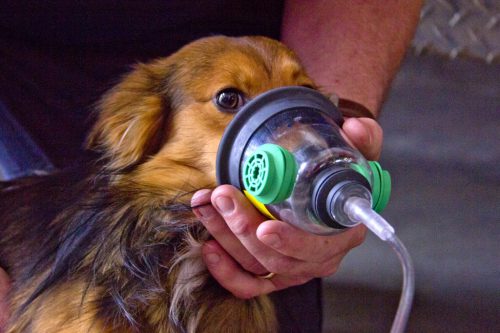
loc-0424-cs-belding-pet-masks-11-1024x6821

This phenomenon of treating numbers without forethought has been one of my pet peeves for years. Somehow, it is assumed that an oximetry value less than the standard “normal” requires therapy. This is not the case.

In young, healthy people the peripheral oxygen saturation values (O2 sat) are typically 96-100% on room air. As we age, the normal values slowly decline. If we abuse ourselves (smoking, working in toxic environments, etc), lung damage occurs and the values can be significantly lower. Patients with obstructive sleep apnea will have much lower numbers intermittently through the night.

So when does a trauma inpatient actually need supplemental oxygen? Unfortunately, the literature provides little guidance on what “normal” really is in older or less healthy patients. Probably because there is no norm. The key is that the patient must need oxygen therapy.

But how can you tell? Examine them! Talk to them! If the only abnormal finding is patient annoyance due to the persistent beeping of the machine, they don’t need oxygen. If they feel anxious, short of breath, or have new onset tachycardia, they probably do. Saturations in the low 90s or even upper 80s can be normal for the elderly and smokers.

Bottom line: Don’t get into the habit of treating numbers without thinking about them. There are lots of reasons for the oximeter to read artificially low. There are also many reasons for patients to have a low O2 sat reading which is not physiologically significant. So listen, talk, touch and observe. Set the alarm level to 90%, or even lower. And if your patient is comfortable and has no idea that their O2 sat is low, turn off the oxygen and toss the oximeter out the window.

Treating Numbers: Pulse Oximetry

How many times has this happened to you? You walk into a young, healthy trauma patient’s room and discover that they have nasal prongs and oxygen in place. Or better yet, these items appear overnight on a patient who never needed them previously. And the reason? The pulse oximeter reading had been low at some point.

This phenomenon of treating numbers without forethought has become one of my pet peeves. Somehow, it is assumed that an oximetry value less than the standard “normal” requires therapy. This is not the case.

In young, healthy people the peripheral oxygen saturation values (O2 sat) are typically 96-100% on room air. As we age, the normal values slowly decline. If we abuse ourselves (smoking, working in toxic environments, etc), lung damage occurs and the values can be significantly lower. Patients with obstructive sleep apnea will have much lower numbers intermittently through the night. 

So when does a trauma inpatient actually need supplemental oxygen? Unfortunately, the literature provides little guidance on what “normal” really is in older or less healthy patients. Probably because there is no norm. The key is that the patient must need oxygen therapy. How can you tell? Examine them! Talk to them! If the only abnormal finding is patient annoyance due to the persistent beeping of the machine, they don’t need oxygen. If they feel anxious, short of breath, or have new onset tachycardia, they probably do. Saturations in the low 90s or even upper 80s can be normal for the elderly and smokers.

Bottom line: Don’t get into the habit of treating numbers without thinking about them. There are lots of reasons for the oximeter to read artificially low. There are also many reasons for patients to have a low O2 sat reading which is not physiologically significant. So listen, talk, touch and observe. If your patient is comfortable and has no idea that their O2 sat is low, turn off the oxygen and toss the oximeter out the window. 

Treating Numbers: Pulse Oximetry

How many times has this happened to you? You walk into a young, healthy trauma patient’s room and discover that they have nasal prongs and oxygen in place. Or better yet, these items appear overnight on a patient who never needed them previously. And the reason? The pulse oximeter reading had been low at some point.

This phenomenon of treating numbers without forethought has become one of my pet peeves. Somehow, it is assumed that an oximetry value less than the standard “normal” requires therapy. This is not the case.

In young, healthy people the peripheral oxygen saturation values (O2 sat) are typically 96-100% on room air. As we age, the normal values slowly decline. If we abuse ourselves (smoking, working in toxic environments, etc), lung damage occurs and the values can be significantly lower. Patients with obstructive sleep apnea will have much lower numbers intermittently through the night. 

So when does a trauma inpatient actually need supplemental oxygen? Unfortunately, the literature provides little guidance on what “normal” really is in older or less healthy patients. Probably because there is no norm. The key is that the patient must need oxygen therapy. How can you tell? Examine them! Talk to them! If the only abnormal finding is patient annoyance due to the persistent beeping of the machine, they don’t need oxygen. If they feel anxious, short of breath, or have new onset tachycardia, they probably do. Saturations in the low 90s or even upper 80s can be normal for the elderly and smokers.

Bottom line: Don’t get into the habit of treating numbers without thinking about them. There are lots of reasons for the oximeter to read artificially low. There are also many reasons for patients to have a low O2 sat reading which is not physiologically significant. So listen, talk, touch and observe. If your patient is comfortable and has no idea that their O2 sat is low, turn off the oxygen and toss the oximeter out the window. 

Pneumothorax And Oxygen: The Final Post!

Okay, this is the last time I’m going to write about this. Hopefully I can provide the final nail in the coffin for this idea. Previously, the oldest paper I could find that was cited as a reason to use high inspired oxygen to treat pneumothorax was from 1983. I found what I think is the earliest (and the last that I will discuss) from 1971!

Twelve patients were retrospectively reviewed who recovered without intervention from a spontaneous pneumothorax. Another 10 were monitored prospectively with the same condition, but were given “high concentration oxygen” (??) by mask from 9 to 38 hours at a time. During intervening periods, the patients breathed room air. Daily chest xrays were obtained, and here is the cool part:

The inner edge of the chest wall and the outer edge of the lung were traced on transparent paper. This was then superimposed on graduated graph paper and the area corresponding to the pneumothorax cavity was measured. The rate of absorption was expressed in cm2/24 hrs.

Need I say more? The authors did show graphically that the apparent rate of absorption tripled in the treated patients, from about 5cm2/day to about 15cm2/day, and was higher in patients with a larger pneumothorax. The problem here is the same as before: chest xray does not allow volumetric estimates, so any results relying on them are suspect. At least it’s not a rabbit study.

Bottom line: There’s just no convincing data to support this practice, so let’s stop using it. Simple physics suggests that this should work, but the effect is just not clinically significant enough to offset the possibility of mishaps from an inpatient admission for oxygen therapy (see yesterday’s post). As I mentioned yesterday, look at the clinical status of your patient. If they have any detectable blood in their chest, they’ll probably need drainage. If not, and if they feel normal, discharge and follow up with a repeat xray in a week. The pneumo will probably be gone. If they do have some compromise, then insert the smallest tube you can. If done properly and a one-way valve can be used, the patient may still be managed as an outpatient.

Related posts:

Reference: Oxygen therapy for spontaneous pneumothorax. Br Med J 4:86-88, 9 Oct 1971.

More On Treating Pneumothorax With Oxygen

One of my readers has pointed out that, yes, the evidence for using O2 to treat pneumothorax is poor, but practice and standard of care are not always driven by evidence. He also pointed out that it’s not really fair to condemn the use of this modality if there isn’t specific evidence showing that it’s bad. In other words, doing something that seems benign is okay if we can’t show that it’s harmful or at least prove that it’s actually benign. I don’t agree.

My point is that no intervention is truly benign. There are always potential complications for the things we do as physicians, sometimes physical, sometimes psychological. Putting a patient on O2 seems safe. But if used as a treatment for pneumothorax, it means hospitalization (which costs a lot of money), an IV (which could get infected), exposure to a lot of sick people (read MRSA and other fun bugs), lying in bed a lot more than at home (DVT), and on and on.

If the pneumothorax does not interfere with function and the patient has decent pulmonary health, why not send them home with reassurance and get a followup chest xray at some point to confirm resolution? If it does cause physiologic problems, or they have pulmonary disease and are likely to develop complications such as pneumonia, then admit for the least invasive treatment to quickly get it out (pigtail type catheter).

Since this topic just won’t seem to die, I’m going to try to kill the last papers I’m aware of on this topic today and tomorrow. Today’s was published in a pediatric surgical journal (!), and it’s another rabbit study. This one adds a wrinkle to the one I discussed yesterday. Not only did they inject air to create a pneumothorax (20cc this time), they punctured the pleura with a needle to create an air leak to simulate a real clinical problem.

They saw the same trend as posted yesterday, although the times were longer. Once again, resolution was measured with chest xray (performed every 12 hours this time). Unfortunately, 7 of the 27 rabbits used in each group died, leaving only 6 or 7 in each of 3 groups for analysis (room air, 40%, 60% O2). Even with wide standard deviations, the authors claimed significant differences in recovery.

Same problems as yesterday, particularly with how resolution of pneumothorax is determined. And don’t use rabbits! A bigger issue is that this is not really a clinically relevant model. First, creating an air leak would defeat the overall purpose of giving high O2 concentrations. If 60% O2 leaked into the pleural space, there would be less nitrogen to wash out so one would think that resolution would take longer. And no one would consider treating a patient with an air leak without some type of drainage device for fear of a tension pneumothorax.

Bottom line: Still not enough evidence to support this seemingly benign treatment. Tomorrow I’ll look at the (hopefully) last paper on the topic since the beginning of time, published in 1971.

Related posts:

Reference: Supplemental oxygen improves resolution of injury-induced pneumothorax. J Pediatric Surg 35(6):998-1001, 2000.

Thanks to Jonathan St. George for his comments on yesterday’s post!