Tag Archives: bladder

4 Guidelines For the Management Of Bladder Injury

The Eastern Association for the Surgery of Trauma (EAST) has been at the forefront of trauma practice guideline dissemination for decades. They recently published a set of recommendations for managing patients with bladder injury. These injuries are not commonly encountered by trauma professionals, and I thought a refresher on current thinking on their management was in order.

Using the usual methodology, the trauma literature was scanned for papers dealing with this topic. After screening for quality, the field was narrowed to 17 papers which were used to formulate the published recommendations. These cover imaging and management questions that frequently come up during the evaluation of these patients.

Following are the questions raised, the EAST recommendations, and my commentary about them:

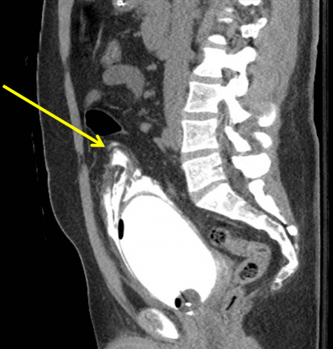
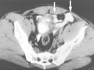
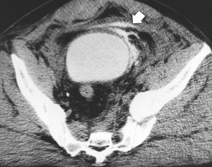
    • In patients with abdominal / pelvic trauma, should retrograde CT cystography vs no imaging be used to diagnose a bladder injury? This seems like a silly question, but the answer lies in the details. It all boils down to the likelihood of injury. And how does one determine likelihood? By looking at the urine and the fracture patterns around the bladder. Patients with microscopic hematuria are very unlikely to have a bladder injury, and any type of bladder imaging in these patients (cystogram, CT cystogram) is almost never positive, and so is not indicated. This is the reason that ordering a urinalysis in major trauma patients is not recommended. However, if gross hematuria is present, CT cystogram is recommended. The sensitivity and specificity are nearly perfect. Just be sure to do a true cystogram by actively filling the bladder with contrast via a urinary catheter. Passive filling of the bladder with urine from the IV contrast misses about half of all the injuries. Also, strongly consider adding CT cystogram in patients with widening of the pubic symphysis. This injury pattern is frequently associated with bladder injury.
    • In patients with intraperitoneal bladder rupture from blunt trauma, should operative or nonoperative management be used to decrease complications? Another silly question? In general, intraperitoneal bladder ruptures do not heal on their own, so urine continues to bathe the peritoneal cavity until the injury is fixed. The review article recommended that operative repair be performed in all of these cases. 
    • In patients with extraperitoneal bladder rupture from blunt trauma, should operative or nonoperative management be used to decrease complications? Patients with a simple extraperitoneal bladder injury should undergo nonoperative management. These injuries usually heal and seal within about 10 days. However, patients with this type of bladder injury that is more complicated (bone spicules piercing the bladder, concomitant vaginal or rectal injury, bladder neck injury) should undergo operative repair in order to decrease the complication rate. One additional group that should be repaired: patients with pubic diastasis that will require operative fixation. The bladder should be repaired at the time of the orthopedic procedure to avoid bathing the new hardware in urine.
    • In patients who have undergone operative or nonoperative management of bladder injury, should bladder closure be assessed with cystogram or not? This one depends on the type and complexity of injury. For simple intraperitoneal bladder injuries that were operatively repaired, no followup cystogram is required. More complex repairs should be evaluated by cystogram before removing the urinary catheter. Finally, simple extraperitoneal injuries should also have a cystogram obtained before removing the catheter. My magic number for obtaining followup studies is 10 days. There is no real science behind this, and no one has systematically looked at 5 vs 7 vs 10 vs 14 days. This one is based only on personal experience.

And by the way, most simple bladder injuries (both intra- and extra-peritoneal) can be easily repaired using two layers by your friendly neighborhood trauma surgeon. More complex injuries are generally best left to the urologist.

Reference: Management of blunt force bladder injuries: A practice management guideline from the Eastern Association for the Surgery of Trauma. J Trauma 86(2):326-336, 2019.

How To: Manage Extraperitoneal Bladder Rupture

Extraperitoneal bladder rupture is a relatively uncommon injury, but is easily managed in most cases. It is associated with a blunt mechanism, and concomitant fracture of the pubic rami or spreading of the symphysis pubis is nearly always present. In the old days, we used to think that the bladder injury was due to penetration anteriorly by bony fragments, but this is probably an old wives tale. It’s more likely due to hydraulic forces occurring within the bladder at the same time the pelvic ring is being deformed or spread apart by blunt forces.

If you obtain a pelvic x-ray during the initial trauma evaluation and see any fractures or diastasis around the symphysis, think bladder injury. Placement of a urinary catheter will typically drain plenty of urine, which will usually be grossly bloody.

Once the injury is suspected, the diagnostic test of choice is a CT cystogram. Don’t confuse this with the images seen when the bladder passively fills with contrast when the catheter is clamped. There is not enough pressure in the bladder to guarantee that contrast will leak out, so this type of study may be falsely negative.

True CT cystogram technique requires filling the bladder with at least 350cc of dilute contrast under pressure by hanging it on an IV pole, then clamping the catheter. Once the bladder is filled, the scan can proceed as usual. But after it is complete, a second limited scan through the pelvis must be performed after the contrast has been evacuated by unclamping the catheter. This allows visualization of small contrast leaks that might otherwise be masked by all the contrast in the bladder.

Here’s a nice sagittal image of an extraperitoneal injury from radiologypics.com:

Note how the contrast dissects around the bladder but does not enter the peritoneal cavity.

Extraperitoneal injuries usually do not require repair and will heal on their own. However, if the symphysis pubis needs instrumentation to restore anatomic position, concomitant repair of the bladder is frequently necessary to keep the hardware from being contaminated by urine.

Bottom line:

  • Suspect an extraperitoneal bladder injury in anyone with bony injuries involving the symphysis pubis.
  • Don’t order a urinalysis in trauma patients!
  • Use CT cystogram technique to make the diagnosis.
  • Treatment is simple: leave the urinary catheter in place for 10 days. No urology consult is needed.
  • Then repeat the CT cystogram to confirm healing, and remove the catheter.

Related posts:

CT Cystography For Bladder Trauma

Bladder injury after blunt trauma is relatively uncommon, but needs to be identified promptly. Nearly every patient (97%+) with a bladder injury will have hematuria that is visible to the naked eye. This should prompt the trauma professional to obtain a CT of the abdomen/pelvis and a CT cystogram.

The CT of the abdomen and pelvis will identify any renal or ureteral (extremely rare!) source for the hematuria. The CT cystogram will demonstrate a bladder injury, but only if done properly!

During most trauma CT scanning of the abdomen and pelvis, the bladder is allowed to passively fill, either by having no urinary catheter and having the patient hold it, or by clamping the catheter if it is present. Unfortunately, this does not provide enough pressure to demonstrate small intraperitoneal bladder injuries and most extraperitoneal injuries.

The proper technique involves infusing contrast into the bladder through a urinary catheter. At least 350cc of dilute contrast solution must be instilled for proper distension and accurate diagnosis. This can be done prior to the abdominal scan. Once the initial scan has been obtained, the bladder must be emptied and a focused scan of just the bladder should be performed (post-void images). Several papers have shown that this technique is as accurate as conventional retrograde cystography, with 100% sensitivity and specificity for intraperitoneal ruptures. The sensitivity for extraperitoneal injury was slightly less at 93%.

Bottom line: Gross hematuria equals CT of the abdomen/pelvis and a proper CT cystogram, as described above. Don’t try to cheat and passively fill the bladder. You will miss about half of these injuries!

Related posts:

Reference: CT cystography with multiplanar reformation for suspected bladder rupture: experience in 234 cases. Am J Roentgenol 187(5):1296-302, 2006.

Intraperitoneal bladder rupture

Extraperitoneal bladder injury

Evaluation of Hematuria in Blunt Trauma

Hematuria ranges from microscopic to gross. Microscopic means blood that can only be seen with a microscope, and gross means visible to the naked eye. In trauma, we only care about gross hematuria, which ranges from the faintest of pink to the deepest red.

In trauma, gross hematuria is a result of an injury to kidney, ureter or bladder. Blunt injury to the ureter is so rare it’s reportable, so you can pretty much forget that one unless the mechanism is extreme. So you really just need to focus on kidney and bladder.

Any victim of blunt trauma that presents with visible hematuria needs to be evaluated by CT of the abdomen and pelvis with an added CT cystogram. Standard CT technique is done without a urinary catheter, or with the catheter clamped. Only 50% of bladder injuries show up with this technique.

CT cystogram is an add-on to the standard CT, and consists of the administration of contrast into the bladder which is then kept under pressure while the scan is done. Delayed slices through the pelvis after the bladder is depressurized and emptied is routine. Nearly 100% of bladder injuries are detected using this technique.

If the CT shows a renal laceration or hematoma, the patient should be admitted and managed according to your solid organ injury protocol. Kidney injuries fare better that livers and spleens, and only rarely require surgery. If no kidney or bladder injury is seen, the default diagnosis of a renal contusion is the culprit. No treatment is needed, and the patient can be discharged if no other injuries are present. The blood will clear over a few days, but may disappear and reappear a few times in the process. The patient can followup with their primary care physician in a week or two.

Related post:

Evaluation of Hematuria in Blunt Trauma

Hematuria ranges from microscopic to gross. Microscopic means blood that can only be seen with a microscope, and gross means visible to the naked eye. In trauma, we only care about gross hematuria, which ranges from the faintest of pink to the deepest red.

In trauma, gross hematuria is a result of an injury to kidney, ureter or bladder. Blunt injury to the ureter is so rare it’s reportable, so you can pretty much forget that one unless the mechanism is extreme. So you really just need to focus on kidney and bladder.

Any victim of blunt trauma that presents with visible hematuria needs to be evaluated by CT of the abdomen and pelvis with an added CT cystogram. Standard CT technique is done without a urinary catheter, or with the catheter clamped. Only 50% of bladder injuries show up with this technique.

CT cystogram is an add-on to the standard CT, and consists of the administration of contrast into the bladder which is then kept under pressure while the scan is done. Delayed slices through the pelvis after the bladder is depressurized and emptied is routine. Nearly 100% of bladder injuries are detected using this technique.

If the CT shows a renal laceration or hematoma, the patient should be admitted and managed according to your solid organ injury protocol. Kidney injuries fare better that livers and spleens, and only rarely require surgery. If no kidney or bladder injury is seen, the default diagnosis of a renal contusion is the culprit. No treatment is needed, and the patient can be discharged if no other injuries are present. The blood will clear over a few days, but may disappear and reappear a few times in the process. The patient can followup with their primary care physician in a week or two.