Tag Archives: bladder

Extraperitoneal Bladder Rupture

This injury is likely to occur in patients who have a full bladder and sustain anterior pelvic trauma that typically leads to fractures. They generally present with gross hematuria upon placement of the bladder catheter. This should prompt an abdominal CT scan with cystogram technique.

CT cystogram involves pressurizing the bladder with contrast prior to the study. This differs from the usual method of clamping the catheter and allowing the bladder to passively fill. The literature here is clear: failure to use cysto technique will miss 50% of these injuries.

The majority of extraperitoneal bladder injuries can be treated nonoperatively, and probably do not need Urology involvement. The bladder catheter is left in place 10-14 days (we do 10 days), and a repeat cystogram is obtained. If there is no leak, the catheter can be removed. If there is still some leakage, Urology consultation should then be obtained. 

There are a few cases where operative management is required:

  • There is some intraperitoneal component of bladder injury
  • Fixation of the pubic rami is required (bathing the orthopedic hardware with urine is frowned upon)
  • Failure of conservative management

Arrows in the photo show extraperitoneal extravasation of cystogram contrast.

Related posts:

Evaluation of Hematuria in Blunt Trauma

Hematuria ranges from microscopic to gross. Microscopic means blood that can only be seen with a microscope, and gross means visible to the naked eye. In trauma, we only care about gross hematuria, which ranges from the faintest of pink to the deepest red.

In trauma, gross hematuria is a result of an injury to kidney, ureter or bladder. Blunt injury to the ureter is so rare it’s reportable, so you can pretty much forget that one unless the mechanism is extreme. So you really just need to focus on kidney and bladder.

Any victim of blunt trauma that presents with visible hematuria needs to be evaluated by CT of the abdomen and pelvis with an added CT cystogram. Standard CT technique is done without a urinary catheter, or with the catheter clamped. Only 50% of bladder injuries show up with this technique.

CT cystogram is an add-on to the standard CT, and consists of the administration of contrast into the bladder which is then kept under pressure while the scan is done. Delayed slices through the pelvis after the bladder is depressurized and emptied is routine. Nearly 100% of bladder injuries are detected using this technique.

If the CT shows a renal laceration or hematoma, the patient should be admitted and managed according to your solid organ injury protocol. Kidney injuries fare better that livers and spleens, and only rarely require surgery. If no kidney or bladder injury is seen, the default diagnosis of a renal contusion is the culprit. No treatment is needed, and the patient can be discharged if no other injuries are present. The blood will clear over a few days, but may disappear and reappear a few times in the process. The patient can followup with their primary care physician in a week or two.

Initial Management of Blunt Bladder Injury

Bladder injury is uncommon after blunt trauma. It is typically seen after high energy events, most commonly a motor vehicle crash with a lap belt in place. During the initial evaluation, the patient may complain of abdominal pain, but this is not universal. 

FAST results are also inconsistent. Free fluid may be seen, and an irregular bladder outline may also be appreciated. The key to diagnosis is placement of a urinary catheter. Bloody urine is found nearly 100% of the time. 

The character of the bloody urine suggests what type of injury is present. Faint hematuria, primarily shades of pink, is usually associated with renal injury or a bladder contusion. A moderate amount of darkly bloody urine is frequently associated with extraperitoneal bladder injury. A small amount of very dark, bloody urine may mean an intraperitoneal bladder injury. Finally, scant and very dark blood in the catheter suggests a urethral injury or a catheter balloon inflated in the urethra.

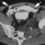
Examination of the urine is suggestive but not diagnostic of the type of injury. Determining the real diagnosis requires imaging, and evaluation of the entire GU tract is essential. CT scan is used to evaluate the kidneys, ureters, and to some degree, the bladder. Cystogram is required to fully evaluate the bladder, and a CT technique may be used. Bladder imaging using passive filling by clamping the catheter is accurate only 50% of the time. The bladder must be pressurized using contrast instilled into the bladder by gravity. When performed in this manner, the CT cystogram is 97% accurate.

Once a diagnosis of bladder injury is made, the treatment is usually straightforward. Extraperitoneal injuries usually do not require repair and will heal on their own. However, if the symphysis pubis needs instrumentation to restore anatomic position, concomitant repair of the bladder is frequently necessary to keep the hardware from being contaminated by urine. 

Intraperitoneal injuries require operative repair. If possible, the injured area should be opened and the inside visually inspected. If the injury extends anywhere near the trigone, a urology consult should be obtained. Most repairs are simple two layer closures. The mucosal layer must be made with absorbable suture; the outer layer is surgeon’s choice. 

For either type of bladder injury, the urinary catheter should be left in place for about 10 days. A cystogram should be obtained, and in most cases there will not be any leakage of urine and the catheter can be removed. In the event of a leak, another 7 days with the catheter is in order and the cystogram can be repeated.

The vast majority of bladder injuries can be easily handled by the trauma surgeon and are healed completely within two weeks.

Related posts: