Galleries

Chest Tube Tips

I’ve written a lot about chest tubes, but there’s actually a lot to know. And a fair amount of misinformation as well. Here’s some info you need to be familiar with:

  • Chest trauma generally means there is some blood in the chest. This has some bearing on which size chest tube you choose. Never assume that there is only pneumothorax based on the chest xray. Clot will plug up small tubes.
  • Chest tubes for trauma only come in two sizes: big (36Fr) and bigger (40Fr). Only these large sizes have a chance in evacuating most of the clot from the pleural space. The only time you should consider a smaller tube, or a pigtail type catheter, is if you know for a fact that there is no blood in the chest. The only way to tell this is with chest CT, which you should not be getting for diagnosis of ordinary chest trauma.
  • When inserting the tube, you have no control of the location the tube goes once you release the instrument used to place it. Some people believe they can direct a tube anteriorly, posteriorly, or anywhere they want. They can’t, and it’s not important (see next tip).
  • Specific tube placement is not important, as long as it goes in the pleural space. Some believe that posterior placement is best for hemothorax, and anterior placement for pneumothorax. It doesn’t really matter because the laws of physics make sure that everything gets sucked out of the chest regardless of position except for things too big to fit in the tube (e.g. the lung).
  • Tunneling the tube tract over a rib is not necessary in most people. In general, we have enough fat on our chest to ensure that the tract will close up immediately when the tube is pulled. A nicely placed dressing is your insurance policy.
  • Adhere to an organized tube management protocol to reduce complications and the time the tube is in the chest.

And finally, amaze your friends! The French system used to size chest tubes is the diameter of the tube in millimeters times three. So a 40Fr chest tube has a diameter of 13.3mm.

Related posts:

Portable CT Scanning For Trauma Patients

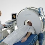
I recently had the opportunity to see a portable head CT scanner in action, the CeroTom by NeuroLogica (Danvers, MA). Today, I’ll give my thoughts on this new technology.

There are 3 major considerations when evaluating portable CT scanning:

  • Patient safety, always at the forefront
  • Usefulness, also know as image quality
  • Financial viability

From a safety standpoint, portable scanning can decrease (but not eliminate) the safety hazards associated with transporting a critically ill patient out of the ICU. Road trips are associated with misplaced/displaced lines, tubes and monitors about 15% of the time. These are lifelines in some patients, and even momentary disruptions can be life-threatening. Some patients are on levels of support so high they are not transportable, so portable scanners offer an opportunity to get diagnostic imaging that would not be available otherwise.

Clinical performance is on par with standard scanners. Resolution is lower, but the diagnostic accuracy and reliability are not different compared to fixed scanners.

From a financial standpoint, use of the portable scanner works as well. The Cleveland Clinic deployed a CereTom scanner a few years ago and found that the unit paid for itself in 6.9 months. For you financial types, the internal rate of return was 169% and the 5-year expected economic benefit was $2.6 million.

Bottom line: This new piece of technology offers significant benefits to patients in the ICU who may otherwise not be able to get imaging due to safety reasons. It can also be employed in the OR on anesthetized patients, which can assist with diagnosis in patients with both abdominal injuries requiring immediate operation and concomitant head injury.

Practical notes: The CereTom is an 8-slice scanner with a 25cm field of view. The patient is moved onto a scan board which supports the head while it is moved slightly off the top of the bed to accommodate the scanner. Current scanner cost is $450,000 and attachment packages for hospital beds are $7,000. One CT technologist can operate the unit, which takes about 5 minutes to set up and 15 minutes to scan. All lines, tubes and monitors must be (carefully) moved to the side of the bed so the scanner can fit over the top.  

References:

  • The economic and clinical benefits of portable head/neck CT imaging in the intensive care unit. Radiology Manage 30(2):50-54, 2008.
  • Review of portable CT with assessment of a dedicated head CT scanner. Am J Neuroradiol 30:1630-1636, 2009.

I have no financial interest in Neurologica, Inc.

Pulmonary Contusion After Sports Injury

Most pulmonary contusions occur after diffuse, high energy blunt trauma like a fall or car crash. Occasionally, pulmonary injury can occur after a much more focal injury like an impact during sports. This may occur due to direct impact from another player, or from a rapidly moving object like a ball or puck. Today, I’ll focus on the latter, impact from a fast-moving object.

Typically, the athlete will complain of pain at the area of impact and some degree of breathing impairment. This is usually due to musculoskeletal pain, rib fracture or involuntary splinting. It is possible to develop pneumothorax or hemothorax, especially if a rib is fractured. At some point, hemoptysis may be present. This is pathognomonic for the presence of a pulmonary contusion.

Any athlete with more than mild to moderate pain, or any physical exam findings other than tenderness, should be more fully evaluated. Here are some important tips:

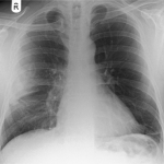
  • The only imaging required is a two view conventional chest xray. This will identify significant pneumothorax, hemothorax or contusions that require additional management.
  • Chest CT is not indicated. It does not change management.
  • Use your typical algorithms for managing hemothorax and pneumothorax
  • A pneumatocele visible on the xray indicates a small pulmonary laceration. A followup xray or two may be needed to ensure that it does not expand or cause a pneumothorax. Thoracic surgery involvement is not usually indicated
  • Hemoptysis is normal, and may present immediately or several days later. The patient should be reassured in advance so they don’t call you frantically in the middle of the night.

Image source: internet

    ACS Review Dinner Tip: Signage

    The ACS Review Dinner is an important part of gathering information on your trauma program. Many of the principal players in your program are present, and the reviewers will have specific questions for most. Although the reviewers may meet many informally before dinner begins, it may be difficult to keep all the names and titles straight.

    Good signs at the table are very helpful. Here are key points on providing the best signage possible:

    • The sign must be large. Compare the size of the sign in the photo to the 8.5×11 inch document next to it.
    • The font size should be large. This allows the reviewers to read name and title from across the room.
    • The same information should be on both sides. Everyone can see their own name so they know where to sit without turning every sign around.

    The sign in the photo is a perfect example of what a good placeholder should look like.

    Where is YOUR Personal Protective Equipment (PPE)?

    Standard or universal precautions are essential in trauma. They serve two purposes: keeping you safe from exposure to body fluids, and keeping you from contaminating any open wounds. Unfortunately, they are not used as “universally” as they should be.

    I’ve heard a number of excuses for not wearing them:

    • I don’t have time to put them on
    • They’re so hot!
    • It’s just a kid, I have nothing to worry about

    All wrong! It takes less than 30 seconds to put them on. And yes, they may be a little warm, but if you have time to notice, then your trauma activations are taking too long. Anyone, including children, may have diseases you don’t want to share.

    There are two major reasons that are legitimate and must be addressed:

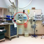
    1. They are not conveniently placed. The deeper in the trauma room they are, the less likely anyone is to wear them (see photo). Place them just outside the door to your trauma bay in plain sight.
    2. Their use is not enforced. Assign specific people the role of PPE police. Emergency physicians and surgeons are optimal, but the charge nurse or others in authority positions are fine.

    Develop a culture where the expectation is that everyone who enters the trauma bay, no matter what their rank, must be wearing their protective gear. Your philosophy should be “it’s not just a good idea, it’s the law.”