Galleries

Technology: The VeinViewer

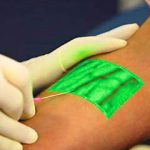
I’m always interested in technology that makes what we do easier. Here’s an objective look at an interesting machine that’s been around for a while. It uses near-infrared light to detect skin temperature changes to allow it to map out veins. It then projects an image of the map in real time onto the skin. In theory, this should make IV starts easier (as long as you can keep your head out of the way of the projector).

A paper just published from Providence, Rhode Island looked at this device to see if it could simplify IV starts in a tertiary pediatric ED. It was a prospective, randomized sample of 323 children from age 0 to 17 looking at time to IV placement, number of attempts, and pain scores.

Unfortunately, the authors did not find any differences. They found that nearly 80% of IVs were started on the first attempt with or without the VeinViewer, which is less than the literature reported 2-3 attempts. This is most likely due to the level of experience of the nurses in this pediatric ED. 

The authors did a planned subgroup analysis of the youngest patients (age 0-2) and found a modest decrease in IV start time (46 seconds) and the nurse’s perception of the child’s pain. Interestingly, the parents did not appreciate a difference in pain between the two groups. This may be due to the VeinViewer’s pretty green display acting as distraction therapy for the child.

Bottom line: This paper points out the importance of carefully reviewing all new (read: expensive at about $20,000 each) technology before blindly implementing it. In this case, an expensive peice of equipment can’t improve upon what an experienced ED nurse can already accomplish.

Reference: VeinViewer-assisted intravenous catheter placement in a pediatric emergency department. Acad Emerg Med, published online, doi: 10.1111/j.1553-2712.2011.01155.x, 2011.

I have no financial interest in Christie Digital Systems, distributor of the VeinViewer Vision®.

Pelvic Trauma Radiographs Demystified

Although we are becoming increasingly reliant on CT scans for diagnosis, plain old radiographs still have their place. This is especially true in pelvic imaging after trauma. 

The most common pelvic radiograph obtained is the supine A-P view taken during trauma resuscitation. This image gives a quick and dirty look at the entire pelvis, from iliac crest to ischial tuberosity. The main areas of interest are the pubic symphysis and the SI joints, so if some of the periphery is cut off a repeat is not necessary prior to CT scan. This image helps predict the need for blood and pelvic compression devices.

If fractures are present, the orthopedic surgeons will generally request additional views in addition to the CT scan. The scan gives excellent detail, but the axial image slices are still not as good as a plain old radiograph in many cases.

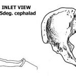
Inlet and outlet views are used to get a better look at the pelvic ring. The inlet view opens the ring up into a big circle (or oval) and allows identification of fractures of the sacrum or displacement of the SI joints, as well as changes in the pubic symphysis. The outlet view shows any vertical displacements through the sacrum or SI joints well, and gives a better appreciation of some pubic fractures.

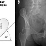
Judet views help demonstrate acetabular fractures by lining up the iliac wing with the xray tube. They can give additional information that the orthopedists use for determining operative or nonoperative management.

Rule of thumb: For major trauma patients, obtain an A-P pelvis radiograph if indicated by mechanism of injury or physical exam. Perform CT scan of the abdomen and pelvis if indicated. If a pelvic ring fracture is identified, obtain inlet and outlet radiographs before calling your orthopedic surgeon. If an acetabular fracture is seen, obtain Judet views before calling.

Compression Of The Fractured Pelvis With A Sheet

Fractures of the posterior pelvis are notorious for their potential to bleed. Here are some tips to use if you encounter a trauma patient with an unstable pelvis and want to slow down the bleeding in the ED.

First, figure out what type of pelvic fracture it is. You will probably be able to do this using physical exam and a simple A-P radiograph. Push down hard on the anterior superior iliac spines to see if the pelvis moves. If so, the patient has an anterior-posterior compression type fracture, and you will likely see diastasis of the pubic bones on the xray. These are amenable to compression maneuvers discussed here.

If the pelvis collapses with lateral compression of the iliac wings, then the patient has a lateral compression fracture and compression maneuvers should not be used. Similarly, if a vertical shear is seen on the xray, do not use compression maneuvers.

There are several pieces of equipment available to help compress the pelvis:

  • Commercial pelvic compression product (e.g. T-Pod). These are convenient but pricey.
  • MAST trousers – just inflate the abdominal compartment, not the legs. But who has these laying around any more?
  • Sheet – cheap and quick. Very effective if used properly.

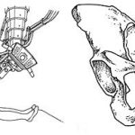
To apply a sheet, it needs to be folded into a narrow band no more than 12 inches high. It should be passed under the patient’s legs and moved upwards. It must be centered over the greater trochanters. This will apply proper pressure, but will not cover the lower abdomen (think laparotomy) or the genitalia (think urinary catheter). Cross the ends of the sheet over as shown above, with one person holding the cinch point while the sheet is secured. This can be carried out with a knot or plastic clamps. Metal clamps will degrade CT or angiographic imaging and should not be used. The sheet should be left in place for the shortest period of time possible, as skin breakdown can occur.

The picture above on the left shows a sheet that is folded too wide (difficult to get enough tension, and covers the good stuff) and uses metal towel clips. The picture on the right shows the proper technique.

Orbital Compartment Syndrome

Sure, you’ve heard about all the other compartment syndromes: leg, thigh, forearm, buttock, and abdomen to name a few. But how about a compartment syndrome of the orbit?

This isn’t your usual muscular compartment problem, although the basic concept is the same. The eye is surrounded by rigid bones or relatively stiff soft tissue (the eyelid, believe it or not). Any extra tissue or blood added to this compartment dramatically raises the pressure in the area, which is readily conducted to the eye itself. This rapidly results in:

  • Severe eye pain
  • Decreased extraocular movements, which may result in diplopia
  • Proptosis
  • Decreased visual acuity
  • Increased intra-ocular pressure (>40 torr)
  • Slow pupillary response

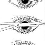
This syndrome should be confirmed rapidly and is one of the few true ophthalmologic emergencies. A lateral canthotomy and cantholysis should be carried out to make the lower lid freely mobile, decompressing the compartment (see diagram). This procedure is not for the faint of heart, but should be familiar in any ED where an ophthalmologist is not readily available. Urgent followup with an eye specialist is mandatory.

Trauma 20 Years Ago: CAVR For Hypothermia

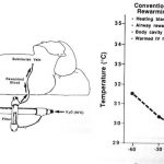
Hypothermia is the bane of major trauma resuscitation, causing mortality to skyrocket. A number of rewarming techniques have been developed over the years. These are classified as passive (the patient generates their own heat) or active (we deliver calories to them), and noninvasive vs invasive. Rewarming speed increases as we move from passive to active and from noninvasive to invasive.

Continuous arteriovenous rewarming (CAVR) is one of the invasive techniques used today. Its use in humans was first reported 20 years ago this month. Larry Gentilello at Harborview in Seattle had experimented with this technique in animals, and reported one case of use in a human who had crashed his car into icy water. After a 20 minute extrication, the patient was pulseless with fixed and dilated pupils, but he regained pulse and blood pressure at the hospital.

The initial core temperature was 31.5C. Peritoneal, bladder and gastric lavage were carried out for warming, as was delivery of warm inspired gas via the ventilator. However, after an hour the temperature had dropped to 29.5C. CAVR was initiated as a last-ditch effort using a jerry-rigged Rapid Fluid Warmer from Level 1 Technologies. The core temperature was raised to 35C after 85 minutes.

The patient did have typical complications (ARDS, acute renal failure), but survived with recovery of his renal and pulmonary function, and a normal neurologic exam. At the time, the authors were unsure whether the complications were due to the near-drowning or the rapid rewarming.

Reference: Continuous arteriovenous rewarming: report of a new technique fo9r treating hypothermia. J Trauma 31(8):1151-1154, 1991.

Related posts: