Galleries

Animal Strikes

This is a bad time of year in much of the United States for striking animals on the road. Car vs animal can be challenging, and motorcycle vs animal is frequently deadly. What can our patients do to protect themselves?

  • Be especially vigilant when driving for the first few hours after sunset and just before sunrise. More animal activity occurs during these hours.
  • If one animal is spotted, look out for others.
  • Drive with high beams on as much as possible. In many animals, this will show reflections from their eyes. Some large animals, such as moose, don’t have glowing eyes.
  • Always where a seat belt in case an impact does occur.
  • If an animal is spotted, slow down quickly and blow the horn.

Most important! NEVER swerve or attempt to quickly change direction. This is one of the most common errors that results in serious injury or death. The driver swerves to avoid and begins to leave the roadway. They then over-correct in the opposite direction and begin a rollover. Always make gentle corrections, staying in the same lane.

For small animals, try to straddle them with the wheels. For larger ones, try to plan the impact so it is in front of the unoccupied front passenger seat. If occupied, plan the strike in the middle of the hood. The idea is to keep the car occupants safe, but to assist with natural selection and remove the animal from the gene pool.

Needle Thoracostomy: Where To Put It?

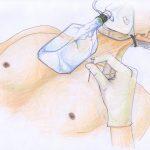
This is another one of those “everything you know is wrong” posts. Since forever, we’ve been taught that an emergent needle thoracostomy should be placed in the second intercostal space, mid-clavicular line. But how do we know?

Once again, the crew at USC+LAC has taken a new look at something we take for granted. They studied thoracostomy insertion in 20 cadavers, using both the classic insertion site as well as a fifth intercostal space, mid-axillary line position.

They found that only 58% of classically placed needles entered the chest cavity, while 100% of the 5th intercostal space catheters were successful. The success rate in the classic position in males was 75%, but in females was only 17%. The authors speculate that the perfect success rate with the lateral approach was due to the absence of extra tissue over the second intercostal space (pectoralis muscle, breast tissue).

Bottom line: Always question dogma. Granted, there are some limitations with this study (using dead people, age and weight not available). Nevertheless, this correlates with my experience, especially when shorter (5cm long) catheters are used. Although I will not necessarily change my practice immediately until there’s a little more literature, I will keep this in mind for obese patients or in those where traditional placement doesn’t seem to be having the desired effect.

Related post:

Reference: Optimal positioning for emergent needle thoracostomy: a cadaver-based study. J Trauma 71(5):1099-1103, 2011.

Using A 3D Printer To Plan Orthopaedic Surgery

I’ve previously written about new printing technology applications in trauma. A recent article details a new way to use 3D printing technology for planning complex orthopaedic procedures.

An orthopedic registrar in Monklands Hospital (North Lanarkshire, Scotland) found a way to combine new printing technology and orthopaedics. CT scans are routinely taken of complex fractures. Scanners now have powerful software that enables us to create 3D reconstructions from the helical or axial images. However, these are just a series of 2D images viewed on a computer monitor.

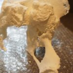
Mr. Mark Frame found a way to convert the CT information into a format that can be used as input for a 3D printer. Using two open source (free) software packages for the Mac, OsiriX and MeshLab, he was able to create a medical quality 3D image file. The file was sent to a company that printed it using a 3D printer.

The cost? About $235 US plus a little time for a complete model of the pelvis. The advantage? The actual size 3D model can be used to select hardware and practice the repair technique. And the cost to own a 3D printer keeps coming down!

Related posts:

There’s An App For That: Reading Xrays on Your iPhone/iPad

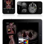
MIM Software has developed an app that allows trauma professionals to view xrays on their mobile device. The app actually received FDA clearance for use earlier this year, and is available as a free download.

The catch is that the software only works with MIM’s cloud-based product for managing radiographic images, called MIMcloud. It is primarily intended for those who do not have access to a full image viewing workstation (MIMfusion).