Tag Archives: TBI

Trauma 20 Years Ago: ED Intubation For Head Injury Is Safe

How far we have come! It’s now commonplace to intubate trauma patients in the ED using rapid sequence induction followed by orotracheal tube placement. However, 20 years ago we were still gnashing our teeth about safety.

In 1991, the group at UMDNJ Newark looked at 100 consecutive trauma patients with suspected head injury who were paralyzed and intubated in the ED. Half of the intubations were performed by a surgeon, the other half by an anesthesiologist. Fifty seven patients were intubated orally and 40 nasally(!). Three required cricothyroidotomy after failure to intubate due to facial fractures.

The majority of these patients had head scans performed; 59% were positive and 15 required emergent neurosurgical procedures. No patients were found to have a neurologic deficit from the intubation even though seven were eventually found to have cervical spine injuries. Only one patient developed an aspiration pneumonia.

The authors concluded that paralysis and intubation in the ED was safe. It helped facilitate the diagnostic workup because they could control combative patients. Up to that time, the only alternative was heavy sedation, which carried its own risks. 

Interesting points on how far we have advanced:

  • Intubation in the ED did not used to be routine. There was a great deal of anxiety before this procedure
  • Nasal intubation was still fairly commonplace
  • The cricothyroidotomy rate was high
  • Intubation was usually performed by a surgeon or anesthesiologist 

Nail Discoloration After Severe Traumatic Brain Injury (TBI)

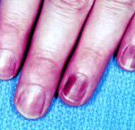
Occasionally, patients who have had a severe brain injury but recovered relatively quickly may present with complaints of odd nail discoloration. This may involve fingernails and/or toenails. What gives?

This is actually a byproduct of repeated exams to determine the Glasgow Coma Scale score. A common way to determine the motor component is to squeeze the fingertip or toetip. I’ve seen some neurosurgeons use a pen to apply a great deal of force to the nail.

The discoloration is a resolving subungual hematoma. You may see different colors under different nails, depending on the age of the hematoma. Amaze your colleagues with your knowledge on this one!

Cervical Spine Clearance and Altered Mental Status

Clearance of the cervical spine is a complicated topic, with many opinions and anecdotes. EAST developed a set of practice guidelines in 1998 and updated them in 2000 and again in 2008. They are well-accepted and very helpful.

Spine clearance in an obtunded or intoxicated patient is made even more challenging. Here’s an approach based on the EAST guidelines that I find helpful:

  1. Clear the bones. Obtain a CT of the cervical spine from skull base to T2. Sagittal and coronal 2D reconstructions must be created for review. Conventional images (AP, lateral, odontoid) are of no additional value.
  2. If a fracture is identified, consult your spine service.
  3. If a neurologic deficit is present, obtain an MRI and consult your neurosurgery or spine service.
  4. Clear the ligaments. In the obtunded patient, there are 3 choices: 1) keep the collar on until the patient wakes up enough to be examined, 2) obtain an MRI to evaluate the ligaments, or 3) remove the collar on the basis of CT alone.

In patients that you don’t expect to wake up any time soon, I prefer MRI. Some say that it should be obtained within 72 hours of injury for best accuracy in detecting ligamentous injury. Unfortunately, I have not been able to find any specific literature support for this. If the MRI is negative, the collar can be removed immediately.

There is a growing body of research that suggests that CT alone is sufficient for clearance. My opinion is that this is probably true, but only if the scan is read by a radiologist who is especially skilled in reading CT scans of the cervical spine. A pool radiologist may miss subtle findings that might indicate a ligamentous injury.

Reference: Eastern Association for the Surgery of Trauma practice guideline: Identifying Cervical Spine Injuries Following Trauma – 2009 Update. Click here to download.

Can Fish-Oil Supplements Speed Recovery From TBI?

Overall, omega-3 fatty acids (O3FA) are thought to be an important and beneficial part of our diet. Recently, the Journal of Neurosurgery published an online paper that looked at the potential benefits of fish oil supplementation on recovery from brain injury.

Originally, axonal damage from TBI was thought to occur at the time of impact. Recent research has shown that the injury is really a progressive event that leads to swelling and axon disconnection during the hours to days after the initial injury. Building on a few animal studies over the past 6 years, a project to look at the effect of omega-3 fatty acid supplementation on brain injury was developed.

The authors performed a controlled study in rats, comparing supplementation with 10 or 40 mg/kg/day of O3FA for 30 days after brain injury with no supplementation. After 30 days, the rats were sacrificed and their brains were examined. The investigators found that a chemical marker of axonal injury (beta amyloid precursor protein) was very significantly decreased in the supplemented animals. The decrease was fairly dramatic and was similar for both doses. 

The actual mechanism by which the protective effect of O3FA was not determined in this study. There is speculation that it may be due to stabilization of brain cell membranes and reduction in the number of reactive oxygen molecules.

This research is very intriguing and appears to have been designed and executed well. The only downside to the work is that the senior investigator is the founder and trustee for the Inflammation Research Foundation. The foundation promotes research on the treatment of diseases with nutritional supplements such as fish oil. The Foundation provided the supplements used in this study. Readers must always be careful when interpreting positive data that is funded or supported by an organization that may benefit from positive results. 

Bottom line: Interesting study, and certainly one that should be followed up with human studies. As far as we know, fish oil supplements are relatively safe, so it should be a little easier to move this work along to human studies. It is not sufficient to recommend adding O3FA to the nutritional regimen of head injured patients yet.

Reference: Omega-3 fatty acid supplementation and reduction of traumatic axonal injury in a rodent head injury model Laboratory investigation. Sears et al. J Neurosurg online July 16, 2010.

Epley’s Maneuver For Vertigo After TBI

Some people experience vertigo after suffering a TBI. This may occur because small calcium carbonate crystals that are normally attached to a membrane in the middle ear are dislodged by the trauma. They can then settle within the semicircular canals. When the head is turned or moved, they brush against the sensitive hairs, sending false signals to the brain. This can result in dizziness, nausea and vertigo. 

The Epley maneuvers were designed to move the crystals back out of the semicircular canals, where they can adhere to the membrane again. They consist of a pattern of head movements that should be performed by a trained professional. This is very important because the maneuvers may induce nausea requiring antiemetics. Certain head movements must be limited for a few days after the maneuvers to make sure the crystals stay in position. The overall success rate is about 80%, but on occasion the maneuvers must be repeated for success.

The video demonstrates the basics of the maneuvers. Remember, don’t try this at home by yourself. Seek out a therapist who is experienced with them.