Tag Archives: Subdural hematoma

A Simple Tool To Predict The Need To Operate On A Subdural Hematoma

Trauma centers in the US are seeing lots of elderly patients, and falls are a major mechanism in the patient group. A significant number sustain a traumatic brain injury. Extra-axial bleeding is fairly common, but because of the increased space available inside the skull, the patient may not become overtly symptomatic. 

So what objective criteria can be used to determine if evacuation of a subdural hematoma (SDH)is needed? A study from the University of Manchester in the UK sought to figure this out. They speculated that the size of the lesion and the amount of displacement it caused might be objective enough. So they set out to see if any specific numbers would provide a reliable method.

Here are the factoids:

  • Two neurosurgeons reviewed four years of head CT scans and determined if they should be treated surgically or nonsurgically.
  • Measurements of the maximum thickness of the lesion, its volume, and the degree of midline shift were taken.
  • Reasonable attempts were made to ensure inter-rater reliability.
  • The total pool of scans studied was 483. 44% were judged to need surgical management.
  • Maximum SDH thickness of 10mm or more, or a midline shift of 1mm or more were found to accurately predict 100% of surgical lesions.
  • The best predictor of the need for surgery was midline shift.
  • Adding hematoma thickness did not significantly improve the ROC curve.

Bottom line: This study is somewhat limited because it is the experience of only one hospital, and the number of clinicians involved in decision making is small. It does echo other similar studies, but in my opinion it omits the use of the mental status exam.

Using a lesion thickness of 10mm or shift of 1mm does not necessarily mean the patient needs surgery if there mental status is completely normal. But these criteria can certainly identify a subset of patients who are at risk, and should be monitored very carefully for any deterioration. A change in GCS by even a single point should then send them straight to OR.

Related posts:

Reference: A simple tool to identify elderly patients with a surgically important acute subdural haematoma. Injury 46(1):76-79, 2015.

Subdural Hematomas and Hygromas Simplified

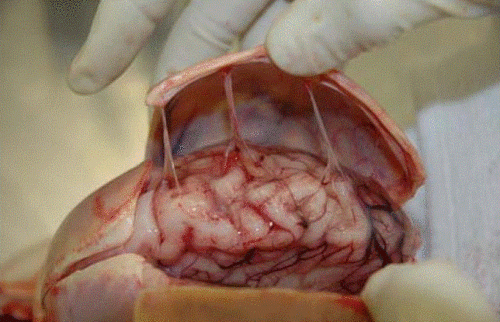
There’s a lot of confusion about subdural pathology after head trauma. All subdural collections are located under the dura, on the surface of the brain. In some way they involve or can involve the bridging veins, which are somewhat fragile and get more so with age.

Head trauma causes a subdural hematoma by tearing some of these bridging veins. Notice how thick the dura is and how delicate the bridging veins are in the image below.

image

When these veins tear, bleeding ensues which layers out over the surface of the brain in that area. If the bleeding does not stop, pressure builds and begins compressing and shifting the brain. A subdural hematoma is considered acute from time of injury until about 3 days later. During this time, it appears more dense than brain tissue.

After about 3-7 days, the clot begins to liquefy and becomes less dense on CT. Many hematomas are reabsorbed, but occasionally there is repeated bleeding from the bridging veins, or the hematoma draws fluid into itself due to the concentration gradient. It can enlarge and begin to cause new symptoms. During this period it is considered subacute.

It moves on to a more chronic stage over the ensuing weeks. The blood cells in it break down completely, and the fluid that is left is generally less dense than the brain underneath it. The image below shows a chronic subdural (arrows).

image

Hygromas are different, in that they are a collection of CSF and not blood. They are caused by a tear in the meninges and allow CSF to accumulate in the subdural space. This can be caused by head trauma as well, and is generally very slow to form. They can lead to slow neurologic deterioration, and are often found on head CT in patients with a history of falls, sometimes in the distant past. CT appearance is similar to a chronic subdural, but the density is the same as CSF, so it should have the same appearance as the fluid in the ventricle on CT.

Related posts:

Subdural Hematoma: How Well Do They Really Do?

The common teaching is that patients with traumatic subdural hematoma don’t do well. This is generally due to the presence of more direct injury to the brain compared with patients who have epidural hematoma. Outcomes data tends to bear this out. However, this data is at least 20 years old and it would be nice to know if we’ve made any progress in the management of this injury.

Harborview Medical Center retrospectively reviewed four years worth of its trauma registry data on patients with subdural hematoma. They scrutinized the usual outcomes data, looking at patients with and without surgical decompression. During the study period, clinical management routines remained basically the same.

A total of 1427 patients were included in the study. The average age was 58. Interesting facts from the study include:

  • Falls were by far the most common mechanism (57%)
  • Most patients (58%) had a GCS of 13 or higher
  • The TRISS probability of survival was slightly lower in the evacuated group (85%) versus the non-evacuated group (91%), yet
  • Mortality rate was 14%, with traumatic brain injury the most common cause of death
  • 29% had positive urine toxicology testing. Marijuana was most prevalent.
  • Slightly more than half were discharged home. Independence was higher in the group who had undergone evacuation of their hematoma.

Bottom line: Patients with subdural hematoma do better these days than they used to. This is probably due to better imaging (CT), which leads to earlier and more accurate management. Additionally, these injuries are now treated at regional trauma centers like Harborview, which may also improve survival.

Related posts:

Reference: Acute traumatic subdural hematoma: Current mortality and functional outcomes in adult patients at a Level I trauma center. J Trauma 73(5):1348-1354, 2012.

Subdural Hematomas and Hygromas Simplified

There’s a lot of confusion about subdural pathology after head trauma. All subdural collections are located under the dura, on the surface of the brain. In some way they involve or can involve the bridging veins, which are somewhat fragile and get more so with age.

Head trauma causes a subdural hematoma by tearing some of these bridging veins. Notice how thick the dura is and how delicate the bridging veins are in the image below.

When these veins tear, bleeding ensues which layers out over the surface of the brain in that area. If the bleeding does not stop, pressure builds and begins compressing and shifting the brain. A subdural hematoma is considered acute from time of injury until about 3 days later. During this time, it appears more dense than brain tissue.

After about 3-7 days, the clot begins to liquefy and becomes less dense on CT. Many hematomas are reabsorbed, but occasionally there is repeated bleeding from the bridging veins, or the hematoma draws fluid into itself due to the concentration gradient. It can enlarge and begin to cause new symptoms. During this period it is considered subacute.

It moves on to a more chronic stage over the ensuing weeks. The blood cells in it break down completely, and the fluid that is left is generally less dense than the brain underneath it. The image below shows a chronic subdural (arrows).

Hygromas are different, in that they are a collection of CSF and not blood. They are caused by a tear in the meninges and allow CSF to accumulate in the subdural space. This can be caused by head trauma as well, and is generally very slow to form. They can lead to slow neurologic deterioration, and are often found on head CT in patients with a history of falls, sometimes in the distant past. CT appearance is similar to a chronic subdural, but the density is the same as CSF, so it should have the same appearance as the fluid in the ventricle on CT.

Related posts: