Tag Archives: spleen

Bedrest After Pediatric Liver/Spleen Injury? Really?

A set of guidelines for management of blunt solid organ injury in children developed by the American Pediatric Surgical Association was originally published in 1999. One of the elements of the guideline was to place the child on bedrest for a period of time after the injury. Arbitrarily, this period was defined as the injury grade plus one day. So for a grade 3 spleen injury, the child would have to stay in bed for 4 days (!).

Recent work looked at the impact of shortening this time interval. Over a 6 year period, all pediatric liver and spleen injuries from blunt trauma were identified and an abbreviated bedrest protocol was implemented. For low grade injuries (grade 1-2), children were kept in bed for 1 day, and for higher grade injuries this was extended to 2 days.

Here are the factoids:

  • 249 patients were enrolled (about 40 per year) with an average age of 10. “Bedrest was applicable for 199 patients, 80%.” Huh? Does that mean that 50 patients were excluded due to surgeon preference?
  • The organ injured was about 50:50 for spleen vs liver. Twelve children injured both.
  • Mean injury grade was 2.7, which is fairly high
  • Mean bedrest was 1.6 days, and mean hospital stay was 2.5
  • Bedrest was the limiting factor for hospital stay in 62% of cases
  • There were no delayed complications of the injury

Bottom line: Come on! Most centers don’t keep adult patients at bedrest this long, and we learned about solid organ injury management from kids! Children almost never fail nonop management, so why treat them more restrictively than adults? And have you ever tried to keep a child at bedrest? Impossible! This study is too underpowered to give real statistically valid results, but it certainly paints a good picture of what works. We’ve been keeping both adults and children at bedrest only overnight, and our average length of stay for isolated solid organs is about 1.5 days. But really, who says that staying in bed for any period of time avoids complications? There are lots of other evil things that can happen!

Related posts:

Reference: Follow up of prospective validation of an abbreviated bedrest protocol in the management of blunt spleen and liver injury in children. J Ped Surg 48(12):2437-2441, 2013.

Contrast Blush in Children

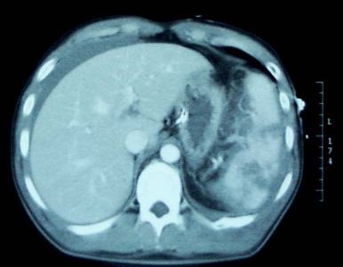
A contrast blush is occasionally seen on abdominal CT in patients with solid organ injury. This represents active arterial extravasation from the injured organ. In most institutions, this is grounds for call interventional radiology to evaluate and possibly embolize the problem. The image below shows a typical blush.

Splenic contrast blush

This thinking is fairly routine and supported by the literature in adults. However, it cannot be generalized to children!

Children have more elastic tissue in their spleen and tend to do better with nonoperative management than adults. The same holds true for contrast blushes. The vast majority of children will stop bleeding on their own, despite the appearance of a large blush. In fact, if children are taken to angiography, it is commonplace for no extravasation to be seen!

Angiography introduces the risk of local complications in the femoral artery as well as more proximal ones. That, coupled with the fact that embolization is rarely needed, should keep any prudent trauma surgeon from ordering the test. A recently released paper confirms these findings.

The only difficult questions is “when is a child no longer a child?” Is there an age cutoff at which the spleen starts acting like an adult and keeps on bleeding? Unfortunately, we don’t know. I recommend that you use the “eyeball test”, and reserve angiography for kids with contrast extravasation who look like adults (size and body habitus).

Reference: What is the significance of contrast “blush” in pediatric blunt splenic trauma? Davies et al. J Pediatric Surg 2010 May; 45(5):916-20.

Financial Impact Of Solid Organ Injury Management

The shift in management of adult solid organ injury from primarily operative to mostly nonoperative began in the late 1980s. For the last decade or so, we’ve been refining this management, figuring out failure criteria, the role of interventional radiology, and developing practice guidelines. We know we’ve been able to reduce the number of people that undergo operative management, with an acceptably low failure rate. But is there a financial impact as well?

Surgeons at the MedStar Hospital Center in Washington DC tapped into a huge hospital discharge database from 1994 to 2010. They focused on patients with admitting diagnoses of spleen or liver injury. They looked at relative costs compared to 1994 practice patterns (still quite a bit of operative management), hospital length of stay, and mortality risk.

Here are the factoids:

  • Nearly 30,000 spleen injury records and 15,000 liver injury records were reviewed
  • Nonop management of spleen injury increased from 38% to 67%, and for liver injury from 62% to 81%
  • In-hospital cost of care decreased by over $8,000 for each patient over the study period
  • Hospital length of stay decreased by about 2 days for each patient
  • Mortality in high risk patients dropped significantly (from 64% to 18% for liver, 30% to 20% for spleen)
  • Mortality in low risk patients remained unchanged (2-3%)

Bottom line: Yes, this study suffers from the usual pitfalls of massaging any large multi-institutional database. But what impresses me is that significant changes have been identified, despite huge variations in how nonoperative management is delivered at so many hospitals. As I have mentioned before, at my hospital we were able to show that just adhering to a standardized solid organ injury protocol squeezes yet another $1000 in costs out of each patient treated, on average. Time to adopt a protocol and adhere to it. Your hospital administrators will love you even more!

Related posts:

Reference: The impact of solid organ injury management on the US healthcare system. J Trauma 77(2):310-314, 2014.

Contrast Blush in Children

A contrast blush is occasionally seen on abdominal CT in patients with solid organ injury. This represents active arterial extravasation from the injured organ. In most institutions, this is grounds for call interventional radiology to evaluate and possibly embolize the problem. The image below shows a typical blush from extravasation.

Splenic contrast blush

This thinking is fairly routine and supported by the literature in adults. However, it cannot be generalized to children!

Children have more elastic tissue in their spleen and tend to do better with nonoperative management than adults. The same holds true for contrast blushes. The vast majority of children will stop bleeding on their own, despite the appearance of a large blush. In fact, if children are taken to angiography, it is commonplace for no extravasation to be seen!

Angiography introduces the risk of local complications in the femoral artery as well as more proximal ones. That, coupled with the fact that embolization is rarely needed, should keep any prudent trauma surgeon from ordering the test. It should be reserved for cases where nonoperative management is failing, but hypotension (hard fail) has not yet occurred.

The only difficult questions is “when is a child no longer a child?” Is there an age cutoff at which the spleen starts acting like an adult and keeps on bleeding? Unfortunately, we don’t know. I recommend that you use the “eyeball test”, and reserve angiography for kids with contrast extravasation who look like adults (size and body habitus).

Reference: What is the significance of contrast “blush” in pediatric blunt splenic trauma? Davies et al. J Pediatric Surg 2010 May; 45(5):916-20.

Complications of Splenic Embolization for Trauma

Angioembolization has become a common procedure that can increase the likelihood of success for nonoperative management for splenic trauma. It does have its own set of complications to be aware of, however.

The most obvious complication is mechanical injury to the femoral artery. This occurs in 1 to 3% of patients. It is more common in the very young (small caliber artery) and the elderly (arteries of stone). Rarely, the substance or device that is used for the embolization may migrate or end up on the wrong spot, infarcting something important.

A common issue that occurs is infarction of portions of the spleen. This is actually the desired effect, as it stops the bleeding. Most of the time, we are unaware of the changes that take place in the spleen post-procedure. But every once in a while we get a repeat CT scan days or weeks down the road and see some very interesting things.

The most common finding is a splenic infarct alone. This is an area of the spleen, sometimes wedge shaped, that does not take up contrast. This is normal. In some cases, gas bubbles are seen within the spleen parenchyma, usually within the infarcted area. In others, large areas of gas are present, and an air-fluid level may also be seen. This is definitely not normal.

Note the infarcted area at the arrow, with a tiny gas bubble visible.

Tiny bubbles are normal after this procedure, and can be ignored if the patient does not appear ill and does not have any systemic evidence of inflammation or sepsis. On the other hand, big bubbles or air-fluid levels probably indicate a developing splenic abscess, and the patient will usually appear ill and have a high WBC count. Unfortunately, the only treatment for this is splenectomy. Insertion of drainage catheters does not work and the patient will only become sicker if it is attempted.