Tag Archives: pop quiz

Pop Quiz: What’s The Diagnosis? The Answer

Okay, time for the answer. This 12 year old crashed his moped, taking handlebar to the mid-epigastrium. Over the next 3 days, he felt progressively worse and finally couldn’t keep food down.

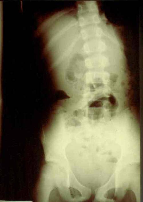
Mom brought him to the ED. The child appeared ill, and had a WBC count of 18,000. The abdomen was firm, with involuntary guarding throughout and a hint of peritonitis. The diagnosis was made on the single abdominal xray shown yesterday. Here is a close-up of the good stuff?
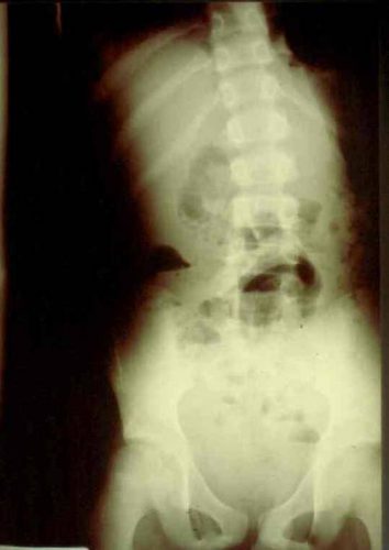
Emergency docs, your differential diagnosis list with this history is a pancreatic vs a duodenal injury based on the mechanism.

Classic findings for duodenal injury:

  • Scoliosis with the concavity to the right. This is caused by psoas muscle irritation and spasm from retroperitoneal soiling by the duodenal leak.
  • Loss of the psoas shadow on the right. Hard to see on this xray, but the left psoas shadow is visible, the right is not. This is due to fluid and inflammation along this plane.
  • Air in the retroperitoneum. In this closeup, you can actually see tiny bubbles of leaked air outlining the right kidney. There are also bubbles along the duodenum and a few along the right psoas.

We fluid resuscitated first (important! dehydration is common and can lead to hemodynamic issues upon induction of anesthesia) and performed a laparotomy. There was a  blowout in the classic position, at the junction of 1st and 2nd portions of the duodenum. The hole was repaired in layers and a pyloric exclusion was performed, with 2 closed drains placed in the area of the leak.

The child did well, and went home after 5 days with the drains out. Feel free to common or leave questions!

Pop Quiz: Do We Really Need To Do All That? The Answer

The scenario involved an elderly woman who fell from standing at her care facility 12 hours earlier. They want to send her to your trauma center for evaluation because she seems a bit different from her baseline. You have well defined practice guidelines for patients with head injuries that dictate what type of monitoring and diagnostics they receive.

What do you need to know to determine what you should do? Thanks for all of you who sent in suggestions.

Here are my thoughts:

  • Which scans should she get? Usually, you would obtain an initial head CT and, due to her age, a cervical CT regardless of her physical exam due to the high miss rate in these patients. But now the fun begins. Your subarachdoid / intraparenchymal hemorrhage (IPH) practice guideline would have you admit for neurologic monitoring for 12 hours, obtain a TBI screen, then discharge without a followup scan if the screen was passed. But in this case, the clock started 12 hours ago and the guideline would be finished with the exception of the TBI screen. So an initial scan and a TBI screen in the ED are all that are needed. The observation period is already over and the patient could potentially be discharged from ED if a SAH or IPH were found.
    Your subdural guideline mandates all of the above plus a repeat scan at 12 hours. But once again, the clock has already started. Do you just get an initial scan, which also serves as the 12 hour scan? Or do you get yet another one?  If the neuro exam is normal, I vote for the former, and your evaluation is complete after the TBI screen. If the neuro exam is not quite normal, then admission for continuing exams and a repeat scan are in order.
  • Does the patient need to be admitted, and for how long? Hopefully, you’ve figure this out in the previous bullet. The clock started running when she fell down, so in cases where the physical exam is normal, only the first CT is needed and ongoing monitoring is not. Thus, she could return to her care facility from the ED after the scan.
  • What other important information do you need to know? Of paramount importance is her DNR status and her/her family’s willingness to have brain surgery if a significant lesion is identified. It is extremely important to know the latter item. If there is never any patient or family intent to proceed to surgery, is there any point to obtaining scans at all? In my opinion, no. The whole reason to obtain the scan and monitor is to potentially “do something.” But if the patient and/or family will not let us “do something,” there is no reason to do any of this. It is crucial that the patient and family understand the typical outcomes from surgery given her age and degree of frailty. This is most important in patients who are impaired with dementia or a high-grade lesion  if found from which there is minimal chance of recovery. In most such cases, even if surgery is “successful,” the patient will never recover enough to return to their prior level of care. This should be weighed heavily by the family and care providers.
  • Should a patient with DNR or “no surgery” orders even be sent to the ED? Theoretically, no. There is no need from the standpoint of their future care. They are not really eligible to have any studies or monitoring done. However, the facility may try to insist for their own liability issues, but this is not really a valid clinical reason.

I hope you enjoyed this little philosophical discussion. Feel free to agree/disagree through your comments or tweets!


What Would You Do? A Teensy Weensy Stab To The Abdomen – Part 4

We’ve gotten the young man with the teensy weensy stab to the abdomen with a bit of omental evisceration to the operating room. Now what should we do? We’ve already decided that he needs an exploration because of the known penetration. How should we go about it?

There are two choices: diagnostic laparoscopy vs laparotomy. Which is better? Let’s talk about laparoscopy first. This tool has been around now for over 25 years. There has been variable acceptance for use in trauma during that time because it tends to take more time and may have a higher rate of missed injury. Both factors have major implications in patients who have active bleeding and small injuries, respectively.

On the plus side, a truly negative (nontherapeutic) exploration tends to be more benign, with rapid recovery, faster time to discharge, and potentially fewer complications when evaluated with a scope. But on the minus side, small injuries can be notoriously difficult to find. What does that small wisp of blood mean? This is not nearly as clear as the meaning of other colors (green, brown). The decision to open can be difficult, particularly for surgeons who perform a high number of laparoscopies in the non-trauma portion of their practice.

Trauma laparotomy is traditionally a large operation with a generous incision and meticulous exploration. This can lead to significant postop pain and morbidity, particularly when no significant pathology is found. Unfortunately, the literature appears to be quite polarized. The surgeon is either pro-laparoscopy, or pro-big incision, and tends to brace their preferred procedure almost exclusively.

But there is a middle ground, and that is what I would choose in a case like this. The surgeon must consider the likelihood of reliably finding the size of internal injury based on his or her assessment of the external wound, as well as the probability that the exploration would be non-therapeutic. So in this case, I would worry that a bowel injury could be only a few millimeters in size and might be missed using only the laparoscope. But I also think that there is a good chance there may not be an injury at all, so I would not be inclined to start with a huge incision.

My choice is to perform a “mini-laparotomy”, making an incision just large enough to explore all of the bowel and visualize the retroperitoneum. I can generally do this through an incision large enough to get my palm into the abdomen, about 6cm. I am confident that I can easily find all injuries, and make the incision larger if warranted. Postoperative pain is better, and discharge if no injuries were found can happen in 1-2 days.

Unfortunately, I can’t find any papers that examine this middle ground between laparoscopy and full laparotomy. But I’ll keep looking! How would you have managed this case? Comment or tweet, please!

In my next post, I’ll review the official algorithm for evaluating stabs to the abdomen recently published by the western Trauma Trauma Association.

References: 

  1. The role of laparoscopy in management of stable patients with
    penetrating abdominal trauma and organ evisceration. J Trauma 81(2):307-311, 2016.
  2. Diagnostic Laparoscopy for Trauma: How Not to Miss Injuries. J Laparoscopic Adv Surg Tech 28(5):506-513, 2018.

What Would You Do? Teensy Weensy Stab To The Abdomen – Part 3

In my last post, I described the plight of a young man who had sustained a stab to the abdomen. It appeared that a very tiny bit of omentum was hanging out of the wound. What to do?

I listed three options:

  • Local wound exploration
  • CT scan of the abdomen
  • Proceed to the operating room

So let’s work through these. First, local wound exploration (LWE).

LWE is a diagnostic procedure to determine if a sharp wound has actually or potentially penetrated a vital area. It is usually performed in the neck to determine if the platysma has been violated, or in the abdomen top check for peritoneal violation. In this case, you would use it if you just couldn’t believe that the bit of odd fat was actually omentum, or if you were unsure what you were looking at. You could also grab it (gently) and give it a little tug. If more comes out, you’ve made your diagnosis. Fortunately, this is rarely necessary because omentum has a very distinctive appearance. You know it when you see it.

What about probing the wound? One of my mentors, John Weigelt, used to ask, “Michael, does your finger / q-tip / instrument have an eyeball on the end of it?” His point was that probing is like so many other medical tests: diagnostic if positive, but unsettling if it’s not. What happens if the wound does penetrate, but you can’t find the path that the knife/bullet took? You can only call that indeterminate. I suppose you could take an approach that includes probing first, then proceeding to full LWE if that is negative.

I’ll describe the proper technique for local wound exploration in a later post.

And what about CT scan? This is another unsatisfying test, because it is very likely to be negative with small wounds. The fascial defect in this case will be very small, and can easily be missed on the scan. Not recommended.

Given all this discussion, my vote is to proceed to the operating room. I know this is omentum, and I know that there is a good likelihood that there will be an injury that needs repair. So let’s go get it done.  But what procedure should I do, and how should I do it? That’s the subject for my next post.

As always, please leave comments below or tweet them out!