Tag Archives: pneumothorax

Is It Really Safe To Observe Occult Pneumothorax?

Occult pneumothorax is the most common incidental finding on CT imaging, occurring in 2% to 10% of trauma patients. By definition, an occult pneumothorax is a pneumothorax that is seen only on CT and not a conventional chest x-ray. When detected, the question that comes to mind is, will this patient need a chest tube?

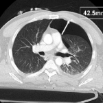
The AAST conducted a trial encompassing the experience at 16 Level I and II trauma centers around the US. They looked at injury severity, specific chest injuries, ventilator settings if on positive pressure ventilation (PPV) and size of pneumothorax. The size was calculated by measuring the largest air collection along a line perpendicular to the chest wall (see image above). Failure of observation meant that a thoracostomy tube was placed.

The 2 year study looked at a total of 448 occult pneumothoraces that were initially observed. Key findings of the study were:

  • Injury severity was no different between failure and non-failure groups
  • There was a 6% failure rate overall
  • PPV alone was associated with an increased failure rate of 14%
  • Surgical intervention requiring PPV was not associated with an increased failure rate
  • Pneumothorax size > 7mm, positive pressure ventilation, progression of the pneumothorax, respiratory distress and presence of hemothorax were associated with failure.
  • Pneumothorax size was not entirely reliable for predicting failure, since patients with sizes as small as 5mm on PPV and 3mm not on PPV failed in this series

Bottom line: Most blunt trauma patients with an occult pneumothorax can be safely observed. A followup chest x-ray should be obtained to look for progression. If the patient progresses, is placed on PPV, has a hemothorax or develops respiratory distress, have a low threshold for inserting a drainage tube. Maximum pneumothorax size may predict failure when large, but it can still happen with very small air collections.

Related posts:

Reference: Blunt traumatic occult pneumothorax: is observation safe? – results of a prospective, AAST multicenter study. J Trauma 70(5):1019-1025, 2011.

CT image courtesy of Journal of Trauma

Pneumothorax in Children

Last week I wrote about pneumomediastinum in children (click here to read). This week I’ll talk about a somewhat more common problem: pneumothorax. This condition is far more mysterious than in adults.

Sports related pneumothorax rarely occurs without rib fractures, which are themselves uncommon in children. The usual mechanism is barotrauma, most likely from an impact while the glottis is closed. The typical presentation is that of pleuritic chest pain, which may be followed by dyspnea. Focal chest wall tenderness is typically absent. Teenagers tend to engage in more strenuous activity and are more likely to actually sustain a rib fracture, so they may have focal tenderness over the fracture site.

Spontaneous pneumothorax in children is also uncommon. However, it is a very different entity. It may be related to blebs in the lungs, and may be more common in children who were born prematurely. The recurrence rate after successful treatment is approximately 50% (in small series). Recurrence is not predictable by looking for blebs on chest CT. The recurrence rate is significantly lower after VATS.

Bottom line: A child who complains of pleuritic chest pain, and especially dyspnea, should undergo a simple PA chest xray. If a pneumothorax is present, consider the following:

  • Insert a small chest tube or catheter if needed, the smaller the better. (I’m stilling looking for the answer to the question of how big a pneumothorax is big enough)
  • Don’t use high inspired oxygen; it doesn’t work. (Read my older blogs from 2010 – this one and this one, too)
  • Don’t get a chest CT for either the initial pneumothorax or any recurrences (too much radiation, too little utility)
  • If this is a spontaneous pneumothorax, caution the parents on the possibility of recurrence
  • If a spontaneous pneumothorax does recur, consult a pediatric surgeon to consider VATS pleurodesis
  • When can the child return to sports? There is absolutely no good literature. I recommend the usual time it takes most soft tissues to return to full tensile strength after injury (6 weeks).

References:

  • Management of primary spontaneous pneumothorax in children. Clin Pediatr, online ahead of print, April 11, 2011.
  • Sports-related pneumothorax in children. Pediatric Emergency Care 21(4): 259-260, 2005.

Related posts:

Thanks to Chris Chow MD for finding some of the literature for this post.

Pneumomediastinum After Falling Down

Finding pneumomediastinum on a chest xray or CT scan always gets one’s attention. However, seeing this condition after a simple fall from standing is very simple to evaluate and manage.

There are 3 potential sources of gas in the mediastinum after trauma:

  • Esophagus
  • Trachea
  • Smaller airways / lung parenchyma

Blunt injury to the esophagus is extremely rare, and probably nonexistent after just falling down. Likewise, a tracheal injury from falling over is unheard of. Both of these injuries are far more common with penetrating trauma.

This leaves the lung and smaller airways within it to consider. They are, by far, the most common sources of pneumomediastinum. The most common pattern is that this injury causes a small pneumothorax, which dissects into the mediastinum over time. On occasion, the leak tracks along the visceral pleura and moves directly to the mediastinum.

Management is simple: a repeat chest xray after 6 hours is needed to show non-progression of any pneumothorax, occult or obvious. This image will usually show that the mediastinal air is diminishing as well. There is no need for the patient to be kept NPO or in bed. Monitor any subjective complaints and if all progresses as expected, they can be discharged after a very brief stay.

Yet Another Paper on Treating Pneumothorax With Oxygen?

Back in February, I thought I closed the door on using high inspired oxygen to try to speed up the resolution of pneumothorax (see related post below). I’ve just run across another attempt which is equally as bad!

This article was published in the Journal of Pediatric Surgery in 2000. The authors randomly divided 27 rabbits into three groups: room air, 40% O2, and 60% O2. Each was given a complete pneumothorax and received chest xrays twice a day. The average time to resolution was measured in each group.

At first glance, it appears that the higher O2 groups resolve faster. But wait, something’s fishy here! In the room air group, the complete pneumothorax went away on its own in 5 days. This doesn’t really happen in people. And in the 60% group, it disappeared in a day and a half! Miraculous!

Oh, and incidentally, a quarter of the rabbits died before completion of the study.

Bottom line: At first glance, these results sure look promising. However, they are rabbits, and they don’t act like people, let alone children! And the resolution times are unrealistic for humans. I still do not recommend the use of high inspired oxygen in an attempt to resolve a pneumothorax. Either some kind of tube is needed for larger volumes (small caliber if air only, bigger if blood is present), or it will go away on its own if the pneumothorax is small.

Related posts:

Reference: Zierold et al. Supplemental oxygen improves resolution of injury-induced pneumothorax. J Pediatric Surg 35(6):998-1001, 2000.

Flying After Pneumothorax

Patients who have sustained a traumatic pneumothorax occasionally ask how soon they can fly in an airplane after they are discharged. What’s the right answer?

The basic problem has to do with Boyle’s Law (remember that from high school?). The volume of a gas varies inversely with the barometric pressure. So the lower the pressure, the larger a volume of gas becomes. Most of us hang out pretty close to sea level, so this is not an issue.

However, flying in a commercial airliner is different. Even though the aircraft may cruise at 30,000+ feet, the inside of the cabin remains considerably lower though not at sea level. Typically, the cabin altitude goes up to about 8,000 to 9,000 feet. Using Boyle’s law, any volume of gas (say, a pneumothorax in your chest), will increase by about a third on a commercial flight. 

The physiologic effect of this increase depends upon the patient. If they are young and fit, they may never know anything is happening. But if they are elderly and/or have a limited pulmonary reserve, it may compromise enough lung function to make them symptomatic.

Commercial guidelines for travel after pneumothorax range from 2-6 weeks. The Aerospace Medical Association published guidelines that state that 2-3 weeks is acceptable. The Orlando Regional Medical Center reviewed the literature and devised a practice guideline that has a single Level 2 recommendation that commercial air travel is safe 2 weeks after resolution of the pneumothorax, and that a chest xray should be obtained immediately prior to travel to confirm resolution.

Bottom line: Patients can safely travel on commercial aircraft 2 weeks after resolution of pneumothorax. Ideally, a chest xray should be obtained shortly before travel to confirm that it is gone. Helicopter travel is okay at any time, since they typically fly at 1,500 feet or less.

References:

  • Practice Guideline, Orlando Regional Medical Center. Air travel following traumatic pneumothorax. October 2009.
  • Medical Guidelines for Airline Travel, 2nd edition. Aerospace Medical Association. Aviation, Space, and Environmental Medicine 74(5) Section II Supplement, May 2003.