Tag Archives: philosophy

Should Extended FAST (eFAST) Be The Standard Of Care In Trauma Activations?

Focused abdominal sonography for trauma (FAST) has been around in one form or another for about 40 years. Sonographic examination of the abdomen was used in Europe in the 1970s, while the US was using diagnostic peritoneal lavage (DPL). FAST finally moved to the US in the 1990s and continues to this day. It has also been incorporated in the Advanced Trauma Life Support Course sponsored by the American College of Surgeons.

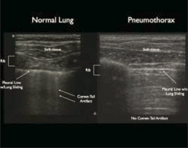
About 10 years ago, emergency physicians began using sonography to evaluate the thorax as well. The technique was primarily used to detect air (and possibly fluid) in the pleural space. Sensitivity and specificity have increased nicely over the years as the technology and our experience has improved.

Most trauma centers incorporate FAST into their trauma activations. Although it was initially vetted using blunt trauma patients, it can be and is used for evaluation in penetrating trauma. But relatively few centers expanded it to eFAST to evaluate the chest. Should they?

Bottom line: Definitely! Extended FAST adds about a minute to the overall exam and may provide information before the chest x-ray is obtained. It may also show pathology that the typical trauma chest x-ray cannot due to patient body habitus and supine positioning. I recommend that the eFAST be the standard of care in trauma activations if you have an ultrasound machine. Important! But be sure to have a way to record and perform quality reviews of the information obtained.

Related posts:

How To Make TEG / ROTEM Useful

A lot of papers have been written on the use of thromboelastography in trauma. And pretty much any meeting or course you may attend has at least one talk on it. And I get it. It can be an important tool in treating trauma patients who have some sort of coagulation disturbance. It helps us figure out what specific part of the coagulation process is out of whack and suggests how we can fix it.

But there are a few problems, as I mentioned yesterday. And the “friction” that those issues cause overall decreases how useful it is. Here’s a partial list of the problems:

  • The equipment costs money, and the disposables that must be used for every patient do, too.
  • Where do you put the machine? Most hospitals can’t put one unit in every possible area it might be used.
  • How to you get the results to a care area if there is no unit there?
  • There is a significant learning curve for interpreting the results
  • How can it be integrated into the massive transfusion protocol?

The main issue is that the current state of TEG and ROTEM are very similar to the state of electrocardiography shortly after it’s discovery. Here’s what you got then:

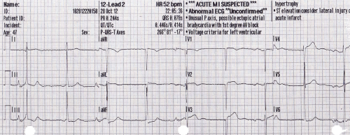
In order to get the most from an EKG, you need to combine this tracing with that from other leads, do a bunch of measurements, look for abnormal shapes and elevations/depressions, etc.  This is exactly where we are with TEG and ROTEM today. Relatively crude, and it takes a lot of work to use it.

The tracing below shows where we are with EKGs today. A computer program looks at all the tracings, and rapidly applies a complex set of rules to come to a set of diagnoses. Notice in the image below that this reading is “unconfirmed.” But how many times in your career have you seen a cardiologist correct one of these? The machines are actually very good!

Bottom line: The tracing above is where we need to be with TEG and ROTEM. Instead of a clinician staring at a developing tracing and figuring out what products to give, these machines need to be just like an automated EKG machine. Sure, a human can still stare at the trace. But the machine will automatically monitor it, apply rules about what abnormalities are present and what is needed to correct them. Send off your blood specimen, and within minutes instructions like “infuse 2 units of plasma now” or “give 12u cryo now” appear. These may be displayed on a monitor in the treatment area, or be broadcast to the phone or pager of the responsible clinicians.

Current TEG/ROTEM equipment is what I would consider 1st generation. The next generation will reduce or remove much of the “friction” in the current process and allow us to really integrate TEG/ROTEM meaningfully into the massive transfusion protocol for trauma. And for anyone who develops this 2nd generation equipment, don’t forget my royalty checks for this idea! 

Related post:

Gunshots And CT Scan Of The Abdomen

Abdominal gunshots and CT scanning are usually thought to be mutually exclusive. The usual algorithm generally means a prompt trip to the operating room. But as with many things in the management of trauma, there are always exceptions. The key is to understand when exactly one of those exceptions is warranted.

Exception 1: Did it really enter the abdomen? Gunshots have enough energy that they usually do get inside. However, freaky combinations of trajectory and body habitus do occur. There are three tests that must be passed in order to entertain the possibility that the bullet may not have made it inside your patient: physiology, anatomy, and physical exam. For physiology, the patient must be completely hemodynamically stable. Anatomically, the trajectory must make sense. If the known wounds and angles allow a tangential course make sense, then fine. But if there is a hole in the epigastrium and another next to the spine, you have to assume the bullet went straight through. Finally, the physical exam must be normal. No peritonitis. No generalized guarding. Focal tenderness only in the immediate area of any wounds. If all three of these criteria are passed, then a CT can be obtained to demonstrate the trajectory.

Exception 2: Did it enter an unimportant area of the abdomen? Well, there’s really only one of these, and that’s the area involving the right lobe of the liver and extending posteriorly and lateral to it. If the bullet hole(s) involve only this area, and the three tests above are passed, CT may confirm an injury that can be observed. However, there should only be a minimal amount of free fluid, and no soft tissue changes of any kind adjacent to bowel.

Exception 3: A prompt trauma lap was performed, but you think you need more information afterwards. This is rare. The usual belief is that the eyes of the surgeon provide the gold standard evaluation during a trauma lap. For most low velocity injuries with an easily understood trajectory, this is probably true. However, high velocity injuries, those involving multiple projectiles, or complicated trajectories (side to side) can be challenging for even the most experienced surgeon. Some areas (think retroperitoneum or deep in the pelvis) are tough to visualize completely, especially when there’s blood everywhere. These are also the cases most likely to require damage control surgery, so once the patient has been temporarily closed, warmed and resuscitated, a quick trip to CT may be helful in revealing unexpected shrapnel, unsuspected injuries, or other issues that may change your management. Even a completely unsurprising scan can provide a higher sense of security.

Bottom line: CT of the abdomen and gunshots to that area may actually coexist in some special cases. Make sure the physiology, anatomy and physical exam criteria are passed first. I also make a point of announcing to all trainees that taking these patients to CT is not the norm, and carefully explain the rationale. Finally, apply the concept of the null hypothesis to this situation. Your null hypothesis should state that your patient does not need a CT after gunshot to the abdomen, and you have to work to prove otherwise!
Practice Guidelines And Tincture Of Time

Most trauma centers have at least a few practice guidelines to help the standardize the way they manage common injuries. Solid organ injury. Elder trauma. Chest tube management. But they are all designed for use in patients who present shortly after their injury.

What about someone who presents a day or two, or more, after their injury?  That changes the picture entirely. Most guidelines have a time component built in. A TBI protocol requires a repeat head CT after a certain period of time. Solid organ injury patients may have restricted activity or frequent vital signs for a while. 

But all too often, trauma professionals treat the patient with delayed presentation exactly the same as fresh trauma. For example, a patient falls and bumps their head. They have a persistent headache, and after two days decide to visit their local ED. The CT scan shows a small amount of subarachnoid blood in the area of the impact. Your practice guidelines says to admit for observation, frequent neruo checks, and repeat head CT in 12 hours.

Or a young male playing sports took a hit to his left flank. After 3 days, he’s just tired of the pain and comes to the ED for some pain medication. CT scan shows a grade III spleen injury with a small amount of hemoperitoneum. Your protocol says to admit, make NPO, liimit activity, and observe for 2 days.

What would I do in these cases? Think about it! If the patients had presented right after the event, they would have gone through your guideline and would have been discharged already. So I would review the images, talk to the patients about their injuries, then send them home from the ED with followup. They’ve already passed!

Bottom line: Remember, practice guidelines are not etched in stone. Variances are possible, but need to be well thought out in advance. And hopefully documented in the chart to expedite the inevitable trauma performance improvement inquiry. If the requisite amount of time has gone by, and the history and exam are reasonable, the patient has already passed your protocol. Send them home.

Related posts:

The Fifth Law Of Trauma – Pediatric

I knew there was a fifth law! Any time I give a pediatric talk, I mention it. This one applies to anyone who takes care of children, and is particularly important to EMS / prehospital providers and emergency physicians.

On occasion, medics are called to a home to treat a child in extremis, or occasionally in arrest. Similarly, extremely sick children are often brought to the ED by parents or other caregivers.

Here’s the fifth law:

A previously healthy child who is in arrest, or nearly so, is a victim of child abuse until proven otherwise.

Bottom line: It’s so easy to go down the sepsis path with sick kids, especially those who can’t talk yet. But healthy children tend to stay healthy, and don’t easily get sick to the point of physiologic collapse. If you encounter one as a prehospital provider, glance around at the environment, and evaluate the caregivers. In the ED, ask pointed questions about the circumstances and do a full body examination. What you hear and what you see may drastically alter how you evaluate the patient and may save their life.

Other Laws of Trauma: