Tag Archives: pelvis

Pelvic Binder Orthosis vs Pelvic External Fixation

Yesterday, I wrote about the open book, A-P compression mechanism, pelvic fracture. In the “old” days, the recommended management for an unstable pelvis like these was application of an external fixator. In some textbooks, it was even suggested that this should be done (by orthopedics) in the resuscitation room. High volume trauma centers with ortho residents could actually pull this off, but not many others.

As the idea of pelvic orthotic binders caught on (T-POD, sheeting, etc) and was adopted by prehospital providers, and then trauma teams, the use of initial external fixation dropped off. But the idea that external fixation was the most desirable or most effective lingered on. A study from Memphis finally sheds some light on the answer to this question.

image

A 10 year retrospective review was carried out on patients presenting with multiple or severe pelvic ring fractures who had early stabilization of the pelvis. Stabilization consisted of external fixation early on, and gradually shifted to pelvic orthotic devices over the study period. They ultimately analyzed outcomes for 93 patients in each treatment group.

The authors found that transfusion needs were dramatically reduced with the orthotic devices (5 units vs 17 units at 24 hours) compared to the orthotics. About a quarter of patients in each group went to angiography, and even in those patients the transfusion need remained lower in the orthotic device group. Hospital length of stay was also significantly shorter in this group (17 vs 24 days). There was no difference in mortality.

Bottom line: Although this is a small, retrospective study it easily showed significant results and will probably never be repeated. Use of a pelvic orthotic device (POD) resulted in less blood replacement and shorter stays in hospital. This technique is simple, cheap and quick, an ideal combination. But does a sheet count as an orthotic device? We don’t know. It’s really cheap, but probably a bit less controlled than a POD. If you have a real POD in your ED or your ambulance, use it. If not, apply the sheet, which will be described tomorrow with other binders.

Tomorrow: what’s the “best” pelvic binder?

Reference: Emergent pelvic fixation in patients with exsanguinating pelvic fractures. JACS 204:935-942, 2007.

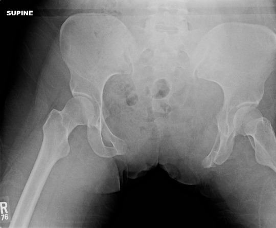
Grading A-P Force Pelvic Injury

Pelvic bony injury requires substantial force, and there are several distinct fracture patterns seen. Today, I’ll briefly review the so called A-P force mechanism and its grading.

The anterior-posterior (A-P) mechanism frequently results in what many call an “open book” pelvis on x-ray evaluation. It most commonly occurs when something heavy rolls over or crushes the pelvis. We see this in patients who have a vehicle roll over their torso, or are crushed by heavy machinery. The force is applied to the sacrum posteriorly and the anterior portions of the iliac crests. This fulcrum effect displaces one or both iliac wings posteriorly. The flexion point is typically the sacro-iliac joint or the sacral wings. The pubic symphysis pulls apart as the iliac wings move away from their anatomic position.

The usual grading system assigns a type subclassification based on the amount of disruption:

  • Type I – less than 1 inch (2.5cm) of pubic diastasis, or rami are fractured; no significant posterior injury
  • Type II – more than 1 inch of diastasis; one or both SI joints widened; posterior SI ligament intact; anterior SI, sacrospinous and sacrotuberous ligaments torn
  • Type III – all anterior and posterior ligaments disrupted

How is this grading system useful? It is generally predictive of hemodynamic instability, resuscitation requirements, and the possibility of concomitant vascular and/or neurologic compromise. However, you can also get a pretty good idea of all of that just looking at the x-ray. But it is helpful in describing the injury to your orthopedic colleagues.

Tomorrow: What to do about it in your trauma bay.

Unstable Patient & Pelvic Fracture + Hemoperitoneum

The usual thinking is that most unstable trauma patients need a quick trip to the OR to stop the bleeding from something. In the US and Europe, patients with nasty pelvic fractures are no exception, especially those with hemoperitoneum. But many of these patients are bleeding from vessels associated with the pelvic fractures and not so much from associated intra-abdominal injuries. And operative management of pelvic fracture bleeding is far from satisfying, even when using preperitoneal packing.

Well, things are a little different in Japan. In many cases, unstable patients are taken to interventional radiology for angio and possible embolization. Is this prudent, or is it dangerous? A Japanese group decided to critically look at this practice by examining the Japan Trauma Data Bank for answers.

Here are the factoids:

  • Patients with pelvic fracture and positive FAST were included, who underwent either laparotomy or angioembolization as their first intervention (n=1153). Those with non-salvageable head injury were excluded, as well as patients who underwent another major procedure first (craniotomy, thoracotomy, ortho procedures, etc.). Only 317 patients remained.
  • In-hospital mortality was the primary outcome of interest
  • A total of 123 underwent laparotomy first, and 194 went to angio first
  • A very small number of patients were hypotensive on arrival (81 laparotomy first, 82 angio first)
  • Half of the patients who were hypotensive on arrival went to angio first (!)
  • Laparotomy-first patients had a higher crude mortality, but this disappeared when confounders were controlled. This was true in patients who were either normotensive or hypotensive on arrival.
  • The authors concluded that the initial intervention should be determined by severity of injury, since in-hospital mortality was no different

Bottom line: Whoa! This is a sweeping statement for a study with so few subjects. Yes, it can be very difficult to determine whether initial bleeding is from the pelvis vs a solid organ or mesenteric injury while in the ED. But it is all too easy to fritter away time (and the patient’s blood/life) in the angiography suite. I recommend trying to stabilize your patient as best you can with fluid and/or blood. If you can maintain a “reasonable” blood pressure, proceed to CT for a quick look at the torso. Then go to the most appropriate location to take care of the problem. And if your patient decompensates in CT or angio, immediately proceed to the operating room!

Related posts:

References:

  • Comparison between laparotomy first versus angiographic embolization first in patients with pelvic fracture and hemoperitoneum: a nationwide observational study from the Japan Trauma Data Bank. Scand J Trauma 21:82, 2013.
  • Eastern Association for the Surgery of Trauma Practice Management Guidelines for Hemorrhage in Pelvic Fracture-Update and Systematic Review. J Trauma 71:1850-1868, 2011.

Pelvic Binder Orthosis vs Pelvic External Fixation

In the “old” days, the recommended management for an unstable pelvis was application of an external fixator. In some textbooks, it was even suggested that this should be done (by orthopedics) in the resuscitation room. High volume trauma centers with ortho residents could actually pull this off, but not many others.

As the idea of pelvic orthotic binders caught on (T-POD, sheeting, etc) and was adopted by prehospital providers, and then trauma teams, the use of initial external fixation dropped off. But the idea that external fixation was the most desirable or most effective lingered on. A study from Memphis finally sheds some light on the answer to this question.

A 10 year retrospective review was carried out on patients presenting with multiple or severe pelvic ring fractures who had early stabilization of the pelvis. Stabilization consisted of external fixation early on, and gradually shifted to pelvic orthotic devices over the study period. They ultimately analyzed outcomes for 93 patients in each treatment group.

The authors found that transfusion needs were dramatically reduced with the orthotic devices (5 units vs 17 units at 24 hours) compared to the orthotics. About a quarter of patients in each group went to angiography, and even in those patients the transfusion need remained lower in the orthotic device group. Hospital length of stay was also significantly shorter in this group (17 vs 24 days). There was no difference in mortality.

Bottom line: Although this is a small, retrospective study it easily showed significant results and will probably never be repeated. Use of a pelvic orthotic device (POD) resulted in less blood replacement and shorter stays in hospital. This technique is simple, cheap and quick, an ideal combination. But does a sheet count as an orthotic device? We don’t know. It’s really cheap, but probably a bit less controlled than a POD. If you have a real POD in your ED or your ambulance, use it. If not, apply the sheet as described below.

Reference: Emergent pelvic fixation in patients with exsanguinating pelvic fractures. JACS 204:935-942, 2007.

What The Heck? The Answer

The photo on Friday shows a woman who had been run over by her own car. The vehicle had rolled over her pelvis and stopped, requiring extrication. The most likely injury is an open book pelvic fracture with significant diastasis and/or bilateral unstable sacral fractures.

If you see this clinical presentation there are several things you need to do immediately:

  1. Call for blood. Losses will be large, so you may even want to consider activating your massive transfusion protocol.
  2. Call an orthopedic surgeon. External stabilization will be needed to help decrease blood loss.
  3. Consider early intubation for control of pain. You will be doing a lot, and a patient in agony will slow you down. Your patient is already hypovolemic, so plan your drug choices accordingly.
  4. Search for evidence of an open fracture. Do a good rectal and vaginal exam looking for blood.

The pelvic xray is poor quality, but shows the major problem, a 10cm pubic diastasis from the open book pelvis fracture. Wrapping the pelvis may be of some help, but consult your orthopedic surgeon first. This pelvis is probably not connected to the spine anymore, so wrapping may have variable results.