Tag Archives: pelvic fracture

Unstable Patient & Pelvic Fracture + Hemoperitoneum

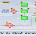
The usual thinking is that most unstable trauma patients need a quick trip to the OR to stop the bleeding from something. In the US and Europe, patients with nasty pelvic fractures are no exception, especially those with hemoperitoneum. But many of these patients are bleeding from vessels associated with the pelvic fractures and not so much from associated intra-abdominal injuries. And operative management of pelvic fracture bleeding is far from satisfying, even when using preperitoneal packing.

Well, things are a little different in Japan. In many cases, unstable patients are taken to interventional radiology for angio and possible embolization. Is this prudent, or is it dangerous? A Japanese group decided to critically look at this practice by examining the Japan Trauma Data Bank for answers.

Here are the factoids:

  • Patients with pelvic fracture and positive FAST were included, who underwent either laparotomy or angioembolization as their first intervention (n=1153). Those with non-salvageable head injury were excluded, as well as patients who underwent another major procedure first (craniotomy, thoracotomy, ortho procedures, etc.). Only 317 patients remained.
  • In-hospital mortality was the primary outcome of interest
  • A total of 123 underwent laparotomy first, and 194 went to angio first
  • A very small number of patients were hypotensive on arrival (81 laparotomy first, 82 angio first)
  • Half of the patients who were hypotensive on arrival went to angio first (!)
  • Laparotomy-first patients had a higher crude mortality, but this disappeared when confounders were controlled. This was true in patients who were either normotensive or hypotensive on arrival.
  • The authors concluded that the initial intervention should be determined by severity of injury, since in-hospital mortality was no different

Bottom line: Whoa! This is a sweeping statement for a study with so few subjects. Yes, it can be very difficult to determine whether initial bleeding is from the pelvis vs a solid organ or mesenteric injury while in the ED. But it is all too easy to fritter away time (and the patient’s blood/life) in the angiography suite. I recommend trying to stabilize your patient as best you can with fluid and/or blood. If you can maintain a “reasonable” blood pressure, proceed to CT for a quick look at the torso. Then go to the most appropriate location to take care of the problem. And if your patient decompensates in CT or angio, immediately proceed to the operating room!

Related posts:

References:

  • Comparison between laparotomy first versus angiographic embolization first in patients with pelvic fracture and hemoperitoneum: a nationwide observational study from the Japan Trauma Data Bank. Scand J Trauma 21:82, 2013.
  • Eastern Association for the Surgery of Trauma Practice Management Guidelines for Hemorrhage in Pelvic Fracture-Update and Systematic Review. J Trauma 71:1850-1868, 2011.

Are Inlet / Outlet Views Obsolete In Pelvic Trauma?

Orthopedic surgeons have long found inlet and outlet views (I/O) of the pelvis to be helpful in their management of pelvic fractures. However, for the last decade we’ve seen an inexorable creep in diagnostic imaging from plain xrays to computed techniques. Have the conventional inlet and outlet views lost their luster?

San Francisco General Hospital and UCSF recently published a registry-based study looking at conventional pelvic I/O images and virtual I/O images reconstructed from CT scans. Two years of registry data were reviewed, and included patients had both conventional I/O images and CT imaging. Images were evaluated by two orthopedic traumatologists for their quality.

Sadly, only 20 patients were available for this study, which makes it an interesting pilot at best. The most interesting results were as follows:

  • Quality of imaging was judged to be equal except when pelvic rotation was present. CT fared better in these cases.
  • Both inlet and outlet views were judged to be better when reconstructed by CT
  • Overall, imaging of all portions of the pelvis was about equal in both types of study
  • The need for repeat studies was identified in nearly half of conventional images, but in only 8% of CT images

Bottom line: CT scanning is slowly becoming the preferred modality for just about any type of trauma imaging. In the 1980’s, head CTs became widespread, followed rapidly by abdominal imaging. Chest CT for definitive diagnosis became commonplace around 2000, and spine imaging by CT has now become the gold standard. Although there are a few throwbacks where conventional imaging has been thought to be better, they are vanishing rapidly. Computing technology can now reconstruct inlet and outlet views of the pelvis, correcting for rotation and angulation in any study of the abdomen/pelvis. And if the reconstructed image is not quite right, the tech can change a few parameters and generate it again and again until the image is perfect. 

Orthopedic surgeons should now expect a nicely formatted set of inlet/outlet CT reconstructions in all trauma patients with pelvic fractures.

Related post:

Reference: Are conventional inlet and outlet radiographs obsolete in the evaluation of pelvis fractures? J Trauma 74(6):1510-1515, 2013.

Pelvic Fractures: OR vs Angio In The Unstable Patient

One of the cardinal rules of trauma care is that hemodynamically unstable patients can only go the the operating room from the ED. No trips to CT, xray, etc. Trauma professionals occasionally try to make exceptions to the rule, but it usually doesn’t work out.

Well, what about the patient with severe pelvic fractures who is or becomes unstable? Pelvic fracture bleeding is not always easy or even possible to control in the OR, and angiography offers a way to identify and stop the bleeding, right?

The trauma group at Ryder in Miami did a lengthy (13 year) retrospective review of their experience with these patients. They looked at every patient who underwent angiography, then identified the subset that went to the OR followed by angiography. There were 134 angio patients and 49 OR to angio patients on whom they based their analysis. Obviously, there is plenty of opportunity for bias in this study, and many of the study patients identified had to be excluded due to incomplete records.

Patients who went to the OR first tended to have similar injury severity but were sicker than the angio alone group. Crystalloid and blood resuscitation volumes were significantly higher in the OR group as well. Most of these patients underwent a laparotomy, and 64% had active intra-abdominal bleeding. None died in OR, and most were left with a damage control abdominal closure.

In the angio group, there were really 2 subsets: angio alone, and angio followed by OR. Mortality in the angio alone group was similar to the OR-angio group. But deaths skyrocketed in those who went from angio to OR (67% vs 20%). This is likely due to them failing angiographic management of bleeding. Three patients died in the angio suite.

Bottom line: There’s a lot of data in this paper, and some of the results can be explained by selection bias. However, they appear to support algorithms released by EAST and the WTA (see diagram above). In general, a trauma patient with severe pelvic fractures and hemodynamic instability needs to go to OR to identify and treat any source of intra-abdominal bleeding. If pelvic bleeding remains a problem, preperitoneal packing may be considered, followed by a trip to angio at that point. The rule that unstable patients should only go to OR (or an ambulance bound for a trauma center if there is no OR) still holds!

Reference: Operating room or angiography suite for hemodynamically unstable pelvic fractures? J Trauma 72(2):364-372, 2012.

Quiz: There is just one extremely rare reason that I know of to move to CT with a hemodynamically unstable trauma patient. Leave a comment with your guess.

Pelvic Trauma Radiographs Demystified

Although we are becoming increasingly reliant on CT scans for diagnosis, plain old radiographs still have their place. This is especially true in pelvic imaging after trauma. 

The most common pelvic radiograph obtained is the supine A-P view taken during trauma resuscitation. This image gives a quick and dirty look at the entire pelvis, from iliac crest to ischial tuberosity. The main areas of interest are the pubic symphysis and the SI joints, so if some of the periphery is cut off a repeat is not necessary prior to CT scan. This image helps predict the need for blood and pelvic compression devices.

If fractures are present, the orthopedic surgeons will generally request additional views in addition to the CT scan. The scan gives excellent detail, but the axial image slices are still not as good as a plain old radiograph in many cases.

Inlet and outlet views are used to get a better look at the pelvic ring. The inlet view opens the ring up into a big circle (or oval) and allows identification of fractures of the sacrum or displacement of the SI joints, as well as changes in the pubic symphysis. The outlet view shows any vertical displacements through the sacrum or SI joints well, and gives a better appreciation of some pubic fractures.

Judet views help demonstrate acetabular fractures by lining up the iliac wing with the xray tube. They can give additional information that the orthopedists use for determining operative or nonoperative management.

Rule of thumb: For major trauma patients, obtain an A-P pelvis radiograph if indicated by mechanism of injury or physical exam. Perform CT scan of the abdomen and pelvis if indicated. If a pelvic ring fracture is identified, obtain inlet and outlet radiographs before calling your orthopedic surgeon. If an acetabular fracture is seen, obtain Judet views before calling.

Compression Of The Fractured Pelvis With A Sheet

Fractures of the posterior pelvis are notorious for their potential to bleed. Here are some tips to use if you encounter a trauma patient with an unstable pelvis and want to slow down the bleeding in the ED.

First, figure out what type of pelvic fracture it is. You will probably be able to do this using physical exam and a simple A-P radiograph. Push down hard on the anterior superior iliac spines to see if the pelvis moves. If so, the patient has an anterior-posterior compression type fracture, and you will likely see diastasis of the pubic bones on the xray. These are amenable to compression maneuvers discussed here.

If the pelvis collapses with lateral compression of the iliac wings, then the patient has a lateral compression fracture and compression maneuvers should not be used. Similarly, if a vertical shear is seen on the xray, do not use compression maneuvers.

There are several pieces of equipment available to help compress the pelvis:

  • Commercial pelvic compression product (e.g. T-Pod). These are convenient but pricey.
  • MAST trousers – just inflate the abdominal compartment, not the legs. But who has these laying around any more?
  • Sheet – cheap and quick. Very effective if used properly.

To apply a sheet, it needs to be folded into a narrow band no more than 12 inches high. It should be passed under the patient’s legs and moved upwards. It must be centered over the greater trochanters. This will apply proper pressure, but will not cover the lower abdomen (think laparotomy) or the genitalia (think urinary catheter). Cross the ends of the sheet over as shown above, with one person holding the cinch point while the sheet is secured. This can be carried out with a knot or plastic clamps. Metal clamps will degrade CT or angiographic imaging and should not be used. The sheet should be left in place for the shortest period of time possible, as skin breakdown can occur.

The picture above on the left shows a sheet that is folded too wide (difficult to get enough tension, and covers the good stuff) and uses metal towel clips. The picture on the right shows the proper technique.