Tag Archives: orthopedics

Using A 3D Printer To Plan Orthopaedic Surgery

I’ve previously written about new printing technology applications in trauma. A recent article details a new way to use 3D printing technology for planning complex orthopaedic procedures.

An orthopedic registrar in Monklands Hospital (North Lanarkshire, Scotland) found a way to combine new printing technology and orthopaedics. CT scans are routinely taken of complex fractures. Scanners now have powerful software that enables us to create 3D reconstructions from the helical or axial images. However, these are just a series of 2D images viewed on a computer monitor.

Mr. Mark Frame found a way to convert the CT information into a format that can be used as input for a 3D printer. Using two open source (free) software packages for the Mac, OsiriX and MeshLab, he was able to create a medical quality 3D image file. The file was sent to a company that printed it using a 3D printer.

The cost? About $235 US plus a little time for a complete model of the pelvis. The advantage? The actual size 3D model can be used to select hardware and practice the repair technique. And the cost to own a 3D printer keeps coming down!

Related posts:

The Medical Orthopaedic Trauma Service

Our population is aging, and falls continue to be a leading cause of injury and morbidity in the elderly. Unfortunately, many elders have significant medical conditions that make them more likely to suffer unfortunate complications from their injuries and the procedures that repair them.

A few hospitals around the world are applying a more multidisciplinary approach than the traditional model. One example is the Medical Orthopaedic Trauma Service (MOTS) at New York-Presbyterian Hospital/Weill Cornell Medical Center. Any elderly patient who has suffered a fracture is seen in the ED by both an emergency physician and a hospitalist from the MOTS team. Once in the hospital, the hospitalist and orthopaedic surgeon try to determine the reason for the fall, assess for risk factors such as osteoporosis, provide comprehensive medical management, provide pain control, and of course, fix the fracture. 

This medical center recently published a paper looking at their success with this model. They retrospectively reviewed 306 patients with femur fractures involving the greater trochanter. They looked at complications, length of stay, readmission rate and post-discharge mortality. No change in length of stay was noted, but there were significantly fewer complications, specifically catheter associated urinary tract infections and arrhythmias. The readmission rate was somewhat shorter in the MOTS group, but did not quite achieve significance with regression analysis.

Bottom line: This type of multidisciplinary approach to these fragile patients makes sense. Hospitalists, especially those with geriatric experience, can have a significant impact on the safety and outcomes of these patients. But even beyond this, all trauma professionals need to look for and correct the reasons for the fall, not just fix the bones and send our elders home. This responsibility starts in the field with prehospital providers, and continues with hospital through the entire inpatient stay.

Related post:

Tomorrow: How bad is a simple urinary tract infection in the elderly?

Reference: The medical orthopaedic service (MOTS): an innovative multidisciplinary team model that decreases in-hospital complications in patients with hip fractures. J Orthopaedic Trauma, ePub ahead of print, doi: 10.1097/BOT.0b013e3182242678, Aug 27, 2011.

Novel Hip Reduction Technique: The Captain Morgan

I wrote about posterior hip dislocation and how to reduce it using the “standard” technique about 9 months ago. Emergency physicians and orthopedic surgeons at UCSF-Fresno just published their experience with a reduction technique called the Captain Morgan.

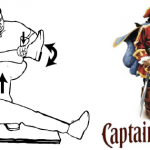
Named after the pose of the trademark pirate for Captain Morgan rum, this technique simplifies the task of pulling the hip back into position. One of the disadvantages of the standard technique is that it takes a fair amount of strength (and patient sedation) to reduce the hip. If the physician is small or the patient is big, the technique may fail.

In the Captain Morgan technique, the patient is left in their usual supine position and the pelvis is fixed to the table using a strap (call your OR to find one). The dislocated hip and the knee are both flexed to 90 degrees. The physician places their foot on the table with their knee behind the patient’s knee. Gentle downward force is placed on the patient’s ankle to keep the knee in flexion, and the physician then pushes down with their own foot, raising their calf. Gentle rotation of the patient’s hip while applying this upward traction behind the patient’s knee usually results in reduction.

Some orthopedic surgeons use a similar technique, but apply downward force on the patient’s ankle, using the leverage across their own knee to develop the reduction force needed. The Captain Morgan technique use the upward lift from their own leg to develop the reduction force. This may be gentler on the patient’s knee.

The authors report a series of 13 reductions, and all but one were successful. The failure occurred due to an intra-articular fragment, and that hip had to be reduced in the operating room. 

I’m interested in hearing comments from anyone who has used this technique (or the leverage one). And does anyone have any other techniques that have worked for them?

Related post:

Reference: The Captain Morgan technique for the reduction of the dislocated hip. Ann Emerg Med (in press) dol:1016/j.annemergmed.2011.07.010, 2011.

Thanks to Sam Stellpflug MD at Regions Hospital for bringing this article to my attention.

Posterior Hip Dislocation

Although posterior hip dislocation is an uncommon injury, the consequences of delayed recognition or treatment can be dire. The majority are caused by head-on car crashes, and 90% of these are posterior dislocations. The femoral head is forced across the back wall of the acetabulum, either by the knee striking the dash, or by forces moving up the leg when the knee is locked. This occurs most commonly on the right side when the driver is standing on the brake pedal, desperately trying to stop.

On exam, the patient presents with the hip flexed, internally rotated and somewhat adducted. Range of motion is limited, and increasing resistance is felt when you try to move it out of position. An AP pelvic X-ray will show the femoral head out of the socket, but it may take a lateral or Judet view to tell if it is posterior vs anterior.

These injuries need to be reduced as soon as possible to decrease the chance of avascular necrosis of the femoral head. Procedural sedation is required for all reductions, since it makes the patient much more comfortable and reduces muscle tone. The ED cart needs to be able to handle both the patient’s weight and your own. I also recommend a spotter on each side of the cart.

Standing on the cart near the patient’s feet, begin to apply traction to the femur and slowly flex the hip to about 90 degrees. Then gently adduct the thigh to help jump the femoral head over the acetabular rim. You will feel a satisfying clunk as the head drops into place. Straighten the leg and keep it adducted. If you are unsuccessful after two tries, there is probably a bony fragment keeping the head out of the socket

Regardless of success, consult your orthopedic surgeon for further instructions. And be sure to thoroughly evaluate the rest of the patient. It takes a lot of energy to cause this injury, and it is flowing through the rest of the patient, breaking other things as well.

Trauma 20 Years Ago: The MESS Score

This month is the 20th anniversary of the MESS score, a system that helps predict salvageability of mangled extremities (Mangled Extremity Scoring System). Obviously, the acronym was chosen to help describe the clinical problem.

The system was originated at the Harborview Medical Center in Seattle. The development was not very scientific; the authors put their heads together and made a list of the four things that they observed predicted limb salvage:

  • Degree of skeletal and soft tissue injury
  • Presence of limb ischemia
  • Presence of shock
  • Age

The system was used retrospectively in a group of 25 patients(!) and the authors found a nice breakpoint at 7. Any mangled extremities with a MESS of 7 or more required amputation. They then applied this to 26 patients prospectively(!) and got the same result.

As you can see, the numbers were small, and there was no followup information. Nevertheless, MESS still stands today, and the critical MESS score has not changed much. It has been validated by a number of other studies during the past 20 years. It is conceivable that the critical score will slowly creep upward with advancements in flap coverage and surgical technique, but it hasn’t done so yet.

Reference: Objective Criteria Accurately Predict Amputation Following Lower Extremity Trauma. Johansen, et al. J Trauma 30(5): 568, 1990.