Tag Archives: IO

The “Double-Barrel” IO: Can It Work?

Intraosseous lines (IO) make life easy. They are quicker to insert, have a higher success rate, and require less experience than a standard IV. And they can be used for pretty much any solution or drug that can be given through an IV.

But there are some limitations. They can’t be inserted into a fractured bone. The manufacturer cautions against multiple insertions into the same bone. A second insertion should not be performed in the same bone within 48 hours. 

But, as with so many things in medicine, there is little in the way of proof for these assertions. They seem like good ideas for precautions, but that does not mean they are correct. No real research has been done in this area. Until now.

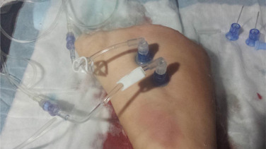
The concept of using two IO needles in one bone was explored in an animal model by researchers in Canada. They used a swine model (using the foreleg/humerus, to be exact), and tested several infusion setups.

Here are the factoids: 

  • Infusing crystalloid using an infusion pump set to 999ml/hr took 30 minutes with a single IO, and 15 minutes with a “double-barrel” setup
  • Giving crystalloid using a pressure bag set at 300 mm/Hg took 24 minutes with a single IO, and 23 minutes with double the fun
  • The double-barrel setup also worked for a blood/drug combo. 250cc of blood and 1 gm of TXA in 100ml of saline infused via pump in 13 minutes.
  • Simultaneous anesthesia drugs (ketamine infusion in IO #1, fentanyl and rocuronium bolus in IO #2) without problems
  • Multiple fluid + drug infusion combinations were tested without incident
  • There were no needle dislodgements, soft tissue injuries, fractures, or macrohistologic damage to the bone or periosteum

Bottom line: Remember, these are pigs. Don’t do this in humans yet. However, this is pretty compelling evidence that the double-barrel IO concept will work in people. And it appears that infusion pumps must be used for effective, fast infusions. I recommend that prehospital agencies with inquiring minds set up a study in people to prove that this works in us, too.

Related posts:

Reference: Double-barrelled resuscitation: A feasibility and simulation study of dual-intraosseous needles into a single humerus. Injury, in press April 30, 2015.

Best Of: CT Contrast Via Intraosseous Catheter

The standard of care in vascular access in trauma patients is the intravenous route. Unfortunately, not all patients have veins that can be quickly accessed by prehospital providers. Introduction of the intraosseous device (IO) has made vascular access in the field much more achievable. And it appears that most fluids and medications can be administered via this route. But what about iodinated contrast agents via IO for CT scanning?

Physicians at Henry Ford Hospital in Detroit published a case report on the use of this route for contrast administration. They treated a pedestrian struck by a car with a lack of IV access sites by IO insertion in the proximal humerus, which took about 30 seconds. They then intubated using rapid sequence induction, with drugs injected through the IO device. They performed full CT scanning using contrast injected through the site using a power injector. Images were excellent, and ultimately the patient received an internal jugular catheter using ultrasound. The IO line was then discontinued.

This paper suggests that the IO line can be used as access for injection of CT contrast if no IV sites are available. Although it is a single human case, a fair amount of studies have been done on animals (goats?). The animal studies show that power injection works adequately with excellent flow rates. 

The authors prefer using an IO placement site in the proximal humerus. This does seem to cause a bit more pain, and takes a little practice. A small xylocaine flush can be administered to reduce injection discomfort in awake patients. Additionally, the arm cannot be raised over the head for the torso portion of the scan. 

Bottom line: CT contrast can be injected into an intraosseous line (IO) with excellent imaging results. Insert the IO in a site that you are comfortable with. I do not recommend power injection at this time. Although the marrow cavity can support it, the connecting tubing may not. Have your radiologist hand-inject and time the scan accordingly.

Note: long term effects of iodinated contrast in the bone marrow are not known. For this reason, and because of smaller marrow cavities, this technique is not suitable for pediatric patients.

Related posts: 

Reference: Intraosseous injection of iodinated computed tomography contrast agent in an adult blunt trauma patient. Annals Emerg Med 57(4):382-386, 2011.

CT Contrast Administration Via Intraosseous Cathether

The standard of care in vascular access in trauma patients is the intravenous route. Unfortunately, not all patients have veins that can be quickly accessed by prehospital providers. Introduction of the intraosseous device (IO) has made vascular access in the field much more achievable. And it appears that most fluids and medications can be administered via this route. But what about iodinated contrast agents via IO for CT scanning?

Physicians at Henry Ford Hospital in Detroit published a case report on the use of this route for contrast administration. They treated a pedestrian struck by a car with a lack of IV access sites by IO insertion in the proximal humerus, which took about 30 seconds. They then intubated using rapid sequence induction, with drugs injected through the IO device. They performed full CT scanning using contrast injected through the site using a power injector. Images were excellent, and ultimately the patient received an internal jugular catheter using ultrasound. The IO line was then discontinued.

This paper suggests that the IO line can be used as access for injection of CT contrast if no IV sites are available. Although it is a single human case, a fair amount of studies have been done on animals (goats?). The animal studies show that power injection works adequately with excellent flow rates.

The authors prefer using an IO placement site in the proximal humerus. This does seem to cause a bit more pain, and takes a little practice see the video above). A small xylocaine flush can be administered to reduce injection discomfort in awake patients. Additionally, the arm cannot be raised over the head for the torso portion of the scan.

Bottom line: CT contrast can be injected into an intraosseous line (IO) with excellent imaging results. Insert the IO in a site that you are comfortable with. I do not recommend power injection at this time. Although the marrow cavity can support it, the connecting tubing may not. Have your radiologist hand-inject and time the scan accordingly.

Note: long term effects of iodinated contrast in the bone marrow are not known. For this reason, and because of smaller marrow cavities, this technique is not suitable for pediatric patients.

Related post: Air embolism from an intraosseous line

Reference: Intraosseous injection of iodinated computed tomography contrast agent in an adult blunt trauma patient. Annals Emerg Med 57(4):382-386, 2011.

CT Contrast Administration Via Intraosseous Cathether

The standard of care in vascular access in trauma patients is the intravenous route. Unfortunately, not all patients have veins that can be quickly accessed by prehospital providers. Introduction of the intraosseous device (IO) has made vascular access in the field much more achievable. And it appears that most fluids and medications can be administered via this route. But what about iodinated contrast agents via IO for CT scanning?

Physicians at Henry Ford Hospital in Detroit have just published a case report on the use of this route for contrast administration. They treated a pedestrian struck by a car with a lack of IV access sites by IO insertion in the proximal humerus, which took about 30 seconds. They then intubated using rapid sequence induction, with drugs injected through the IO device. They performed full CT scanning using contrast injected through the site using a power injector. Images were excellent, and ultimately the patient received an internal jugular catheter using ultrasound. The IO line was then discontinued.

This paper suggests that the IO line can be used as access for injection of CT contrast if no IV sites are available. Although it is a single human case, a fair amount of studies have been done on animals (goats?). The animal studies show that power injection works adequately with excellent flow rates. 

The authors prefer using an IO placement site in the proximal humerus. This does seem to cause a bit more pain, and takes a little practice. A small xylocaine flush can be administered to reduce injection discomfort in awake patients. Additionally, the arm cannot be raised over the head for the torso portion of the scan. 

Bottom line: CT contrast can be injected into an intraosseous line (IO) with excellent imaging results. Insert the IO in a site that you are comfortable with. I do not recommend power injection at this time. Although the marrow cavity can support it, the connecting tubing may not. Have your radiologist hand-inject and time the scan accordingly.

Note: long term effects of iodinated contrast in the bone marrow are not known. For this reason, and because of smaller marrow cavities, this technique is not suitable for pediatric patients.

Related post: Air embolism from an intraosseous line

Reference: Intraosseous injection of iodinated computed tomography contrast agent in an adult blunt trauma patient. Annals Emerg Med 57(4):382-386, 2011.

Air Embolism From an Intraosseous (IO) Line

IO lines are a godsend when we are faced with a patient who desperately needs access but has no veins. The tibia is generally easy to locate and the landmarks for insertion are straightforward. They are so easy to insert and use, we sometimes “set it and forget it”, in the words of infomercial guru Ron Popeil.

But complications are possible. The most common is an insertion “miss”, where the fluid then infuses into the knee joint or soft tissues of the leg. Problems can also arise when the tibia is fractured, leading to leakage into the soft tissues. Infection is extremely rare.

This photo shows the inferior vena cava of a patient with bilateral IO line insertions (black bubble at the top of the round IVC). During transport, one line was inadvertently disconnected and probably entrained some air. There was no adverse clinical effect, but if the problem is not recognized and the line closed, there could be.