Tag Archives: facial fracture

The Tripod Fracture

The tripod fracture (officially known as the zygomaticomaxillary complex fracture, and sometimes called a malar fracture) is the most common one seen after trauma. Fundamentally, the zygoma is separated from the rest of the face in a tripod fracture.

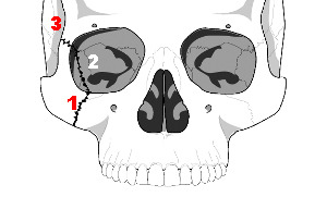
As you might imagine (tripod fracture), there are three components to this fracture. The first is a fracture through the zygomatic arch (1). Next, the fracture extends across the floor of the orbit and includes the maxillary sinus (2). Finally, the fracture includes the lateral orbital rim and wall (3). 

Extraocular muscles may become trapped in the fracture line, leading to diplopia. It is very important to do a good eye exam to try to detect entrapment. The infraorbital nerve also passes through the orbital floor and may be injured, leading to numbness along the lower eyelid and upper lip.

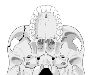
Nondisplaced fractures are treated symptomatically and reevaluated after a week or so to see if surgery would be beneficial. Displaced or symptomatic fractures require early open reduction. The pictures below show the anatomy of these fractures. They are derived from teaching materials provided by the Radiology Department at the University of Washington.
image

image

The Tripod Fracture

The tripod fracture (officially known as the zygomaticomaxillary complex fracture, and sometimes called a malar fracture) is the most common one seen after trauma. Fundamentally, the zygoma is separated from the rest of the face in a tripod fracture.

As you might imagine (tripod fracture), there are three components to this fracture. The first is a fracture through the zygomatic arch (1). Next, the fracture extends across the floor of the orbit and includes the maxillary sinus (2). Finally, the fracture includes the lateral orbital rim and wall (3). 

Extraocular muscles may become trapped in the fracture line, leading to diplopia. It is very important to do a good eye exam to try to detect entrapment. The infraorbital nerve also passes through the orbital floor and may be injured, leading to numbness along the lower eyelid and upper lip.

Nondisplaced fractures are treated symptomatically and reevaluated after a week or so to see if surgery would be beneficial. Displaced or symptomatic fractures require early open reduction. The pictures below show the anatomy of these fractures. They are derived from teaching materials provided by the Radiology Department at the University of Washington.

Diagnosing Facial Fractures With CT

Facial fractures are common after major blunt trauma. There are a number of diagnostic tests available for their diagnosis, including head CT, conventional facial imaging and facial CT.

Our preference has been to add a facial CT to the list of diagnostics in any patient with external evidence of facial trauma. Subjectively, it appeared that there were not many injuries being identified, and the vast majority did not require operative management. 

A review of the literature shows that head CT alone is sufficient for screening for significant facial fractures. A small retrospective series noted that the accuracy was 92%, with 90% sensitivity and 95% specificity. 

Bottom line: A head CT alone ordered for the usual indications is a very good screening test for facial fractures. If none are seen, it is unlikely that there are any fractures that require specific management. If fractures are seen, consultation with a facial surgeon is needed. However, unless the fractures involve critical areas (e.g. temporal bone near the middle ear) or are significantly displaced, the benefit of a facial CT scan is still very low since most will be treated without operation.

Reference: Computed tomography of the head as a screening examination for facial fractures. Marinaro et al. Am J Emerg Med 25, 616-619, 2007.