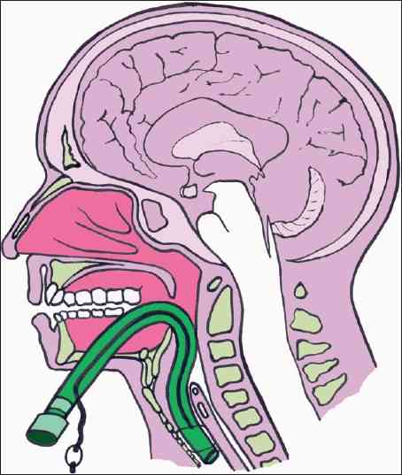
Category Archives: Technique

By Request: Submental Intubation

I keep getting requests regarding this technique, so I’m reposting  this updated article today and tomorrow.

Here’s one of the weirder procedures I’ve seen in some time. Imagine that you need a definitive airway, but you can’t use the face for some reason (mouth or nose). The usual choice would be a tracheostomy, right? But what if you only need it for a few days? Typically, once placed, trachs must be kept for a few weeks before decannulation is safe.

Enter submental intubation. This technique involves passing an endotracheal tube through the anterior floor of the mouth, and then down the airway. This leaves the facial bones, mandible, and skull base untouched.

tumblr_inline_mvds1c14ye1qa4rug1


The technique is straightforward:

  • After initially intubating the patient  orotracheally, a 1.5cm incision is created just off the midline in the submental area under the chin.
  • Using a hemostat, all layers are penetrated, entering the oropharynx just lateral to the tongue.
  • A 1.5cm incision is then made at the puncture site, parallel to the gum line of the lower teeth.
  • The ET tube is removed from the ventilator circuit, and the connector at the proximal end of the tube is removed.
  • The hemostat is placed through the chin incision again. The proximal end of the ET tube is curled into the oropharynx and grasped with the hemostat, then pulled out through the skin under the chin, leaving the distal (balloon) end in the trachea.
  • The connector is reinserted, and the tube is then hooked up to the anesthesia circuit again.
  • The tube is then secured using a stitch under the chin.

After a final position check, the surgical procedure can commence. Cool!


There are a number of variations on this technique, so you may encounter slightly different descriptions. The tube can be pulled at the end of the procedure, or left for a few days to ensure safe extubation, if needed.

tumblr_inline_mvdsm0zd8b1qa4rug1

A small series of 10 patients undergoing this technique was reviewed, and there were no short or long term problems. Scarring under the chin was acceptable, and was probably less noticeable than a trach scar.

Bottom line: This is a unique and creative method for intubating patients with very short-term airway needs while their facial fractures are being fixed. Brilliant idea!

Tomorrow: Submental intubation – the video!

Reference: Submental intubation in patients with panfacial fractures: a prospective study. Indian J Anaesth 55(3):299-304, 2011.

Photo source: internet

Print Friendly, PDF & Email

Update: The Rectal Exam In Trauma Continues to “Pass”?

This topic continues to come up from time to time. I still see trauma programs that perform the good, old-fashioned digital rectal exam on nearly every trauma patient. But is it really necessary?

In the not so distant past, it was standard operating procedure to perform a digital rectal exam in all major trauma patients. The belief had always been that valuable information about blood in the GI tract, the status of the urethra, and the neuro exam (rectal tone) could be gleaned from this exam.

Unfortunately, a finger in the bum also serves to antagonize or even further traumatize some patients, especially those who may be intoxicated to some degree. On a number of occasions I have seen calm patients become so agitated by the rectal exam that they required intubation for control.

So is it really necessary? A study in 2001 conducted over a 6 month period (1) showed that the rectal exam influenced management in only 1.2% of cases. The authors felt that there was some utility in 3 special cases:

  • Spinal cord injury – looking for sacral sparing
  • Pelvic fracture – looking for bone shards protruding into the rectum
  • Penetrating abdominal trauma – looking for gross blood

A more recent 2005 study (2) was also critical of the rectal exam and found that using “other clinical indicators” (physical exam and other diagnostic study information) was at least equivalent, changing management only 4% of the time. They concurred with the first two indications above as well.

And what is the best patient position for the exam? I continue to see people try to do it when the patient is in the logroll position! This is substandard for two reasons:

  • It is not a stable position, and no one likes a finger in their butt. Awake patients will squirm and withdraw, defeating any attempt at spinal precautions.
  • It’s not ideal for the examiner, either.  Access to the male prostate is subpar because the examiner’s finger is generally pointed posteriorly, away from this organ. In order to rotate anteriorly, the examiner must spin around, putting “reverse English” (billiards reference) on their arm.

To do a proper rectal exam when indicated, make sure the patient is supine, warn them that you are going to do it, and use the same hand as the side of the patient you are standing on. Right side, right hand to avoid the “reverse English” thing again.

Bottom line: For most major trauma patients, the rectal exam is not worth the patient aggravation it causes. I still recommend it for the 3 special cases listed above, however, as there are no equivalent and effective exams for these potentially serious patient problems. And remember, DON’T do it while the patient is in the logroll position

References:
1. Porter, Urcic. Am Surg. 2001 May;67(5):438-41.
2. Esposito et al. J Trauma. 2005 Dec;59(6):1314-9.

Print Friendly, PDF & Email

How Fast Can You Warm Up A Hypothermic Patient?

For most places, including Minnesota, hypothermia time is just about over! However, this trauma problem can occur nearly anywhere and at any time. And especially during a massive resuscitation. The optimal way to warm paitients has been debated for years. A number of very interesting techniques have been devised. Ever wonder how fast / effective they are?

I’ve culled data from a number of sources, and here is a summary what I found. And of course, the disclaimer: “your results may vary.”

Warming Technique Rate of Rewarming
Passive external (blankets, lights) 0.5° C / hr
Active external (lights, hot water bottle) 1 – 3° C / hr
Bair Hugger (a 3M product, made in Minnesota of course!) 2.4° C / hr
Hot inspired air in ET tube 1° C / hr
Fluid warmer 2 – 3° C / hr
GI tract irrigation (stomach or colon, 40° C fluid, instill for 10 minutes, then evacuate) 1.5 -3° C / hr
Peritoneal lavage (instill for 20-30 minutes) 1 – 3° C / hr
Thoracic lavage (2 chest tubes, continuous flow) 3° C / hr
Continuous veno-venous rewarming 3° C / hr
Continuous arterio-venous rewarming 4.5° C / hr
Mediastinal lavage (thoracotomy) 8° C / hr
Cardiopulmonary bypass 9° C / hr
Warm water immersion (Hubbard or therapy tank) 20° C / hr
Print Friendly, PDF & Email

The Trauma Activation Pat-Down?

Yes, this is another one of my pet peeves. During a trauma activation, we all strive to adhere to the Advanced Trauma Life Support protocols. Primary survey, secondary survey, etc. Usually, the primary survey is done well.

But then we get to the secondary survey, and things get sloppy.

police_frisk1

The secondary survey is supposed to be a quick yet thorough physical exam, both front and back. But all too often it’s quick, and not so thorough. There is the usual laying on of the hands, but barely. Abdominal palpation is usually done well. But little effort is put into checking stability of the pelvis. The extremities are gently patted down with the hope of finding fractures. Joints are slightly flexed, but not stressed at all.

Is it just a slow degradation of physical exam skills? Is it increasing (and misguided) faith in the utility of the CT scanner? I don’t really know. But it’s real!

Bottom line: Watch yourself and your team as they perform the secondary survey! Your goal is to find all the injuries you can before you go to imaging. This means deep palpation, twisting and trying to bend extremities looking for fractures, stressing joints looking for laxity. And doing a good neuro exam! Don’t let your physical exam skills atrophy! Your patients will thank you.

Print Friendly, PDF & Email

Chest Tube Repositioning – Final Answer

So you’re faced with a chest tube that “someone else” inserted, and the followup chest xray shows that the last drain hole is outside the chest. What to do?

Well, as I mentioned, there is very little written on this topic, just dogma. So here are some practical tips on avoiding or fixing this problem:

  • Don’t let it happen to you! When inserting the tube, make sure that it’s done right! I don’t recommend making large skin incisions just to inspect your work. Most tubes can be inserted through a 2cm incision, but you can’t see into the depths of the wound. There are two tricks:
    • In adults with a reasonable BMI, the last hole is in when the tube markings show 12cm (bigger people need bigger numbers, though)
    • After insertion, get into the habit of running a finger down the radiopaque stripe on the tube all the way to the chest wall. If you don’t feel a hole (which is punched through the stripe), this will confirm that the it is inside, and that the tube actually goes into the chest. You may laugh, but I’ve seen them placed under the scapula. This even looks normal on chest xray!
  • Patients with a high BMI may not need anything done. The soft tissue will probably keep the hole occluded. If there is no air leak, just watch it.
  • If the tube was just put in and the wound has just been prepped and dressed, and the hole is barely outside the rib line, you might consider repositioning it a centimeter or two. Infection is a real concern, so if in doubt, go to the next step.
  • Replace the tube, using a new site. Yes, it’s a nuisance and requires more anesthetic and possibly sedation, but it’s better than treating an empyema in a few days.

Related posts:

Print Friendly, PDF & Email