Category Archives: General

What Is: The LisFranc Injury?

Medicine is full of conditions with eponyms. Trauma is no exception. There’s the Mattox maneuver and the Cushing response, to name two. Many times, the name is just a kind of vanity plate for the discoverer of the condition. But in the case of the LisFranc injury (or fracture), it makes some sense. This injury is tough to describe in a sentence or two, let alone a few words. 

Jacques LisFranc de St. Martin was a French surgeon and gynecologist (!) who described this condition in about 1815. It entails the fracture of the heads of the metatarsal bones and possible dislocation from the tarsals (the cuboid, navicular, and three cuneiform bones). This area is known as the LisFranc joint complex.

image

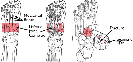
The injury can involve any or all of the metatarsals. The typical mechanism applies high energy across the midfoot, which can often be seen in head-on motor vehicle crashes. Crush injury to the proximal foot can also do this, such as running the foot over with a car. Occasionally, this injury pattern is produced with lower energy during sports play. In this case, the top of the foot is typically contacting the ground, plantar flexing it. At the same time, another player steps on the heel, grinding the foot into the ground (ouch). Interestingly, LisFranc did not describe the injury pattern or mechanism. His name is associated with the joint complex, and it is an injury to his joint complex.

image

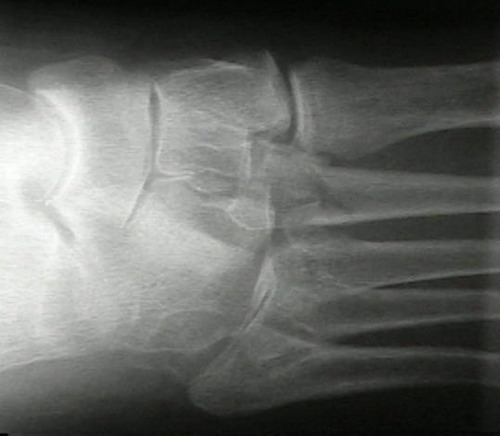
Most of the time, the injury is obvious. There is usually notable pain and swelling of the foot. X-ray findings are generally not subtle. However, lower energy mechanisms may not cause much displacement, and initial imaging may not show the injury. If your patient starts to complain of pain in the midfoot when they begin to ambulate, think of LisFranc.

Treatment depends on the degree of displacement and the amount of disruption of the tarso-metatarsal joints. If minimal, a trial of nonoperative, non-weight bearing may be sufficient. But frequently, surgical reconstruction is required. 

… And The Ultimate Retained Foreign Bodies

Sponges are unfortunately one of the most common retained foreign bodies. This is due to their small, flimsy nature. The surgeon usually looks at the obviously visible areas of the abdomen or other body cavity before closing. She can also feel around in the “nooks and crannies”, but sponges feel very similar to the other organs surrounding it.

But what about more substantial items, like surgical instruments? Surely these are so obvious as to not leave behind?

Unfortunately, not so. Take a look at these items. This is a large pari of surgical forceps.

This is a malleable retractor, a long, thin sheet of pliable metal that can be bent to any desired shape.

And finally, a pair of Metzenbaum scissor, a common surgical instrument for cutting tissue.

Bottom line: It doesn’t matter how small or large, anything can and will be left behind in emergent and trauma cases. Recognizing that this can occur, no matter how confident you are that it has not, is the key. Always count, but followup with an x-ray that covers all areas of the surgical field before closing.

Related post:

What Does A Retained Surgical Sponge Look Like?

It’s the bane of any surgeon’s existence. And the reason why OR personnel take such great pains to account for everything in the room. It is a catastrophe, and always a preventable one, when some piece of equipment goes missing and ends up left inside a patient.

A number of methods have been developed to try to eliminate this problem. They include careful counts, having someone record anytime anything is placed inside, x-rays, and most recently, RFID tags. 

After counting, x-ray is the most common way to try to find missing objects. One would think that these foreign bodies would be easy to see. Metallic instruments are rather easy to spot. But many trauma professionals, even those who work in the OR, have never seen what a positive image of a sponge actually looks like. So here they are. You should never miss one on an xray now.

Surgeons typically use two types of sponges in the OR: Ray-Tec sponges and standard lap pads. Ray-Tecs look like a 4×8 piece of gauze with a mysterious blue string woven throughout it. The string is the only part that shows up on x-ray, and it is very thin and somewhat hard to see. Here are some Ray-Tec sponges outside the body:

And here’s one that was left inside. Note the little squiggle in the left lower quadrant and how easy it is to over look.

On the other hand, a laparotomy pad is a 4×4 folded cloth pad that unfolds into a larger pad. It has a blue tag sewn in the corner, extending along one edge of the pad. Here’s what they look like:

And here’s one inside a patient. Note the irregular object in the right upper quadrant.

Bottom line: It’s important for anyone who works in the OR on any body part to be familiar with the appearance of these tags on x-rays. Since it’s generally impossible to get accurate counts before or after a trauma procedure, always image the involved body cavity looking for these telltale signs before closing the patient.

Note: These images taken from the internet. Patients not treated at Regions Hospital.

September Trauma MedEd Newsletter Released

The September newsletter is now available! Click the image below or the link at the bottom to download. This month’s topic is chest trauma.

In this issue you’ll find articles on:

  • Why I didn’t like finger thoracostomy
  • Advanced needle thoracostomy
  • Trochar vs needle for tension pneumothorax
  • Troubleshooting chest tube air leaks
  • Chest tube collection systems gone wild
  • Managing chest tube air leaks
  • Pneumothorax in children

Subscribers received the newsletter first last weekend. If you want to subscribe (and download back issues), click here.

Click here to download and/or subscribe.

Return To Baseline After Concussion

Here’s another interesting paper that was presented at the Congress of Neurological Surgeons. There’s a lot of attention being focused on the incidence and management of concussion during sporting events. An international Concussion in Sport Group has been meeting for over 10 years, contemplating classification and management of this injury. They are considering using age to modify management of concussion in young athletes.

The authors looked at their own experience with 200 adolescent and young athletes. They stratified by age (younger = 13-16 year olds, older = 18-22 year olds), with 100 in each group. They matched them by number of previous concussions, and all underwent baseline and post-concussion ImPACT testing. They specifically looked at the number of days needed to return to baseline.

Interestingly, they identified significant differences in recovery time. And strangely enough, the older players did better than the younger ones. Overall, 90% returned to baseline within a month. But the younger players took 2-3 days longer to recover than the older ones. 

Bottom line: Looks like the Concussion in Sport Group is right on! Usually in trauma, older folks do worse than younger ones, so we tend to treat them more carefully. Not so in youngsters with concussions. Sports medicine physicians need to realize that the younger brain takes longer to recover, and they should err on the safe side and keep them out of the game longer. Objective testing to help predict return to play is extremely helpful.

Related post:

Reference: Sport-Related Concussion and Age: Number of Days to Neurocognitive Baseline. Oral presentation 145 – Congress of Neurological Surgeons 2012.