Category Archives: General

February Trauma MedEd Newsletter Released

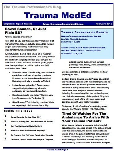
The February newsletter is now available! Click the image below or the link at the bottom to download. This month’s topic is “Tips & Tricks”.

In this issue you’ll find articles on:

  • Bowel sounds, or just plain BS?
  • Tired of waiting for the ambulance to arrive?
  • But the radiologist made me do it!
  • What is a wide mediastinum anyway?
  • To probe or not to probe: penetrating wounds
  • Don’t get lateral view chest x-rays to diagnose pneumothorax

Subscribers received the newsletter first last weekend. If you want to subscribe (and download back issues), click here.

Click here to download the current issue.

Trauma Mortality vs Cancer Mortality from CT Scans for Trauma

Trauma professionals worry about radiation exposure in our patients. A lot. There are a growing number of papers dealing with this topic in the journals every month. The risk of dying from cancer due to CT scanning is negligible compared to the risk from acute injuries in severely injured patients. However, it gets a bit fuzzier when you are looking at risk vs benefit in patients with less severe injuries. Is it possible to quantify this risk to help guide our use of CT scanning in trauma?

A nice paper from the Mayo clinic looked at their scan practices in 642 adult patients (age > 14) over a one year period. They developed dose estimates using a detailed algorithm, and combined them with data from the Biological Effects of Ionizing Radiation VII data. The risk level for injury was estimated using their trauma team activation criteria. High risk patients met their highest level activation criteria, and intermediate risk patients met their intermediate level activation criteria.

Key points in this article were:

  • Average radiation dose was fairly consistent across all age groups (~25mSv)
  • High ISS patients had a significantly higher dose
  • Cumulative risk of cancer death from CT radiation averaged 0.1%
  • This risk decreased with age. It was highest in young patients (< 20 yrs) at 0.2%, and decreased to 0.05% in the elderly (> 60 yrs)

Bottom line: Appropriate CT scan use in trauma evaluation is challenging. It’s use is widespread, and although it changes management it has not decreased trauma mortality. This paper shows that the risk of death from trauma in the elderly outweighs the risk of death from CT scan radiation. However, this gap narrows in younger patients with less serious injuries because of their very low mortality rates. Therefore, we need to focus our efforts to reduce radiation exposure on our young patients with minor injuries.

Related posts:

References:

  • Comparison of trauma mortality and estimated cancer mortality from computed tomography during initial evaluation of intermediate-risk trauma patients. J Trauma 70(6):1362-1365, 2011.
  • Health risks from low levels of ionizing Radiation: BEIR VII, Phase 2. Washington DC: The National Academies Press, 2006.

Serial Hemoglobin / Hematocrit – Huh?

The serial hemoglobin (Hgb) determination. We’ve all done them. Not only trauma professionals, but other in-hospital clinical services as well. But my considered opinion is that they are not of much use. They inflict pain. They wake patients up at inconvenient hours. And they are difficult to interpret. So why do them?

First, what’s the purpose? Are you looking for trends, or for absolute values? In trauma, the most common reason to order is “to monitor for bleeding from that spleen laceration” or some other organ or fracture complex. But is there some absolute number that should trigger an alarm? If so, what is it? The short answer is, there is no such number. Patients start out at a wide range of baseline values, so it’s impossible to know how much blood they’ve lost using an absolute value. And we don’t use a hemoglobin or hematocrit as a failure criterion for solid organ injury anymore, anyway.

What about trends, then? First, you have to understand the usual equilibration curve of Hgb/Hct after acute blood loss. It’s a hyperbolic curve that reaches equilibrium after about 3 days. So even if your patient bled significantly and stopped immediately, their Hgb will drop for the next 72 hours anyway. If you really want to confuse yourself, give a few liters of crystalloid on top of it all. The equilibration curve will become completely uninterpretable!

And how often should these labs be drawn? Every 6 hours (common)? Every 4 hours (still common)? Every 2 hours (extreme)? Draw them frequently enough, and you can guarantee eventual anemia.

Bottom line: Serial hemoglobin/hematocrit determinations are nearly worthless. They cost a lot of money, they disrupt needed rest, and no one really knows what they mean. For that reason, my center does not even make them a part of our solid organ injury protocol. If bleeding is ongoing and significant, we will finding it by looking at vital signs and good old physical exam first. But if you must, be sure to explicitly state what you will do differently at a certain value or trend line. If you can’t do this and stick to it, then you shouldn’t be ordering these tests in the first place!

Related post:

Reference: Serial hemoglobin levels play no significant role in the decision-making process of nonoperative management of blunt splenic trauma. Am Surg 74(9):876-878, 2008.
February Newsletter Released To Subscribers This Weekend!

The February Trauma MedEd Newsletter will be released to subscribers over the weekend. This month’s theme is “tips and tricks.” Articles include:

  • What exactly is a wide mediastinum?
  • Bowel sounds, or just BS?
  • Predicting ambulance arrival time
  • Do you really need to do what the radiologist says?
  • And more!

Anyone on the subscriber list as of 8 PM (CST) Saturday will receive it on Sunday. I’ll release it to everyone else next Friday via the blog. So sign up for early delivery now by clicking here!

Pick up back issues here!

How To Avoid Missed Injuries

I’ve just spent two days here looking at the phenomenon of “delayed diagnosis” or missed injury. I believe that there are only two fundamental reason why this occurs:

  • Insufficient diagnostic technique – A good physical exam and/or specific diagnostic techniques were not performed. Or rarely, the injury cannot be readily detected by existing techniques and technology. The former is usually the real problem, and may be an issue with either the physical exam completeness and/or technique, or judgment used to obtain the appropriate diagnostic test. Example 1: a penetrating injury to the back is missed because the patient is not logrolled to examine this area. Example 2: a spine fracture is missed in an elderly patient with a fall from standing because the back pain found on physical exam is evaluated only with conventional imaging of the spine, not CT.
  • Failure to recognize the injury – The injury was actually identified on a test, but was not appreciated by the clinician. Example 1: the radiologist may not have appreciated and reported out a subtle anomaly in the cervical spine imaging. Example 2: you fail to check you patient’s lab tests and miss a sudden spike in serum amylase or lipase the day after your patient was kicked in the epigastrium by a horse.

So what can you do to avoid this potential problem? Here are some tips:

  • Admit that it can really happen to you. If the missed injury rate at your center is off the low end of the bell curve (< 5%) then you are either really good or really blind. You’d better take a close look at your performance improvement process, because you may be fooling yourself.
  • Adopt a firm definition of “delayed diagnosis.” Basically, you need a time frame after which a new diagnosis is considered “delayed.” It should be a reasonable time interval after the patient has left the ED. If it’s too short an interval (e.g. once they leave the ED), your number will be unnecessarily high. If it’s too long (days and days later), then significant morbidity may occur that you don’t account for. Most centers have adopted 24, 36, or 48 hours after patient arrival.
  • Implement a tertiary survey process. This is a complete physical re-examination followed by a review of all diagnostic studies (lab and radiology) that have been performed. This exam needs to be dated and timed to ensure that it is performed within the time frame noted above. If a new finding is discovered on the tertiary survey, it is not considered a delayed diagnosis. If found after the survey (or after the pre-determined time interval), it is and must be entered into your performance improvement process.
  • Be paranoid. I hate the phrase, “maintain a high index of suspicion” because it’s meaningless. It’s like those stupid “start seeing motorcycles” bumper stickers. You can’t see what you can’t see. But you can be suspicious all the time, constantly looking for the inevitable clinical surprises of trauma care. 

Related posts: