Category Archives: General

Conservative Management Of Pancreatic Injury

There has been a slow shift toward nonoperative management of many injuries that used to demand a quick trip to the operating room. Liver and spleen injury is one of the best examples, with extremely good success rates (95%). Kidneys fall into this category, too.

The pancreas is another solid organ. Perhaps we can do the same thing? A number of pediatric surgeons have been attempting to manage children with pancreatic injury. Low grade injuries (principally contusions) have been managed expectantly for some time. Could higher grade injuries (duct injury) be managed this way as well? How about using repeat imaging, percutaneous drainage, stenting via ERCP, and TPN to avoid the OR in hemodynamically stable kids?

A recent paper looks at this practice critically. Nine years of registry data at two Level I pediatric centers was reviewed to identify all high grade (III-IV) pancreatic injuries. They isolated 39 children with this injury (which is quite a few!). They were separated into two groups based on initial management plan, operative (15) or nonoperative (24). Here are the results of interest (all statistically significant):

  • Average ISS was higher in the nonop group (23 vs 15)
  • Hospital length of stay was longer in the nonop group (28 vs 15 days)
  • TPN was required for a longer period in the nonop group (22 vs 8 days)
  • There were more complications in the nonop group (17 vs 4 children), with 13 developing a pseudocyst (none in the op group)

Bottom line: Nonoperative management of high grade pancreatic injury in kids is just not ready for prime time. It may seem that avoiding a big abdominal operation would be a good thing. Distal pancreatectomy usually keeps children in the hospital for 5-7 days, and then they are done unless they have other serious injuries. Nonoperative management results in a lengthy stay in the hospital, multiple imaging studies (radiation), getting stuck with big drainage needles, and TPN with its attendant infection risks. The old fashioned way, going to the OR, is still the best!

Related post:

Reference: Non-operative management of high-grade pancreatic trauma: is it worth it? J Ped Surg 48:1060-1064, 2013.

Print Friendly, PDF & Email

How Long Are Trauma Patients At Risk For VTE?

Venous thromboembolism (VTE) and its complications are one of the banes of the trauma professional’s existence. Trauma centers have initiated extensive systems of risk assessment, screening, prophylaxis, and treatment of patients at risk for this problem. But typically, much of this management ends at or shortly after discharge from the hospital.

How long do we need to worry? Some trauma programs continue prophylaxis on at-risk patients until they are ambulating well, or for an arbitrary period of time, like one month. But until recently, we’ve had no guidance based on actual numbers. A California study may shed some light on this gray area.

A large dataset from a state of California hospital discharge database was massaged, looking at 6 years of data from patients at the highest risk for VTE (injuries of the pelvis, spine, and spinal cord). The authors looked forward in time after the initial discharge to see if there were any future admissions for VTE and its complications. 

Here are the factoids:

  • Patients with spinal cord injury had the highest risk of VTE, pelvic fractures were mid-range, and vertebral fractures the lowest risk.
  • Occurrence of VTE was associated with a significant risk of mortality, but it was not possible to determine why.
  • In all groups, the risk of VTE remained for the first 3 months after injury, then declined rapidly.
  • VTE risk returned to the level of the general population after about 12 months in patients with pelvic and vertebral injuries.
  • VTE risk in spinal cord injured patients followed a similar curve, but never completely returned to the population baseline.

Bottom line: Obviously, this is not a clinical study. But it’s size and duration is unprecedented and provides valuable information anyway. This information calls into question our existing treatment intervals for prevention of VTE. However, it does not provide real and actionable guidance yet. Additional clinical studies will be needed to parse out the best drugs and duration of treatment.

Related posts:

Reference: Can we ever stop worrying about venous thromboembolism after trauma? J Trauma 78(3):475-481, 2015.

Print Friendly, PDF & Email

February Trauma MedEd Newsletter Released

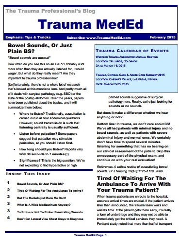
The February newsletter is now available! Click the image below or the link at the bottom to download. This month’s topic is “Tips & Tricks”.

In this issue you’ll find articles on:

  • Bowel sounds, or just plain BS?
  • Tired of waiting for the ambulance to arrive?
  • But the radiologist made me do it!
  • What is a wide mediastinum anyway?
  • To probe or not to probe: penetrating wounds
  • Don’t get lateral view chest x-rays to diagnose pneumothorax

Subscribers received the newsletter first last weekend. If you want to subscribe (and download back issues), click here.

Click here to download the current issue.

Print Friendly, PDF & Email

Trauma Mortality vs Cancer Mortality from CT Scans for Trauma

Trauma professionals worry about radiation exposure in our patients. A lot. There are a growing number of papers dealing with this topic in the journals every month. The risk of dying from cancer due to CT scanning is negligible compared to the risk from acute injuries in severely injured patients. However, it gets a bit fuzzier when you are looking at risk vs benefit in patients with less severe injuries. Is it possible to quantify this risk to help guide our use of CT scanning in trauma?

A nice paper from the Mayo clinic looked at their scan practices in 642 adult patients (age > 14) over a one year period. They developed dose estimates using a detailed algorithm, and combined them with data from the Biological Effects of Ionizing Radiation VII data. The risk level for injury was estimated using their trauma team activation criteria. High risk patients met their highest level activation criteria, and intermediate risk patients met their intermediate level activation criteria.

Key points in this article were:

  • Average radiation dose was fairly consistent across all age groups (~25mSv)
  • High ISS patients had a significantly higher dose
  • Cumulative risk of cancer death from CT radiation averaged 0.1%
  • This risk decreased with age. It was highest in young patients (< 20 yrs) at 0.2%, and decreased to 0.05% in the elderly (> 60 yrs)

Bottom line: Appropriate CT scan use in trauma evaluation is challenging. It’s use is widespread, and although it changes management it has not decreased trauma mortality. This paper shows that the risk of death from trauma in the elderly outweighs the risk of death from CT scan radiation. However, this gap narrows in younger patients with less serious injuries because of their very low mortality rates. Therefore, we need to focus our efforts to reduce radiation exposure on our young patients with minor injuries.

Related posts:

References:

  • Comparison of trauma mortality and estimated cancer mortality from computed tomography during initial evaluation of intermediate-risk trauma patients. J Trauma 70(6):1362-1365, 2011.
  • Health risks from low levels of ionizing Radiation: BEIR VII, Phase 2. Washington DC: The National Academies Press, 2006.
Print Friendly, PDF & Email

Serial Hemoglobin / Hematocrit – Huh?

The serial hemoglobin (Hgb) determination. We’ve all done them. Not only trauma professionals, but other in-hospital clinical services as well. But my considered opinion is that they are not of much use. They inflict pain. They wake patients up at inconvenient hours. And they are difficult to interpret. So why do them?

First, what’s the purpose? Are you looking for trends, or for absolute values? In trauma, the most common reason to order is “to monitor for bleeding from that spleen laceration” or some other organ or fracture complex. But is there some absolute number that should trigger an alarm? If so, what is it? The short answer is, there is no such number. Patients start out at a wide range of baseline values, so it’s impossible to know how much blood they’ve lost using an absolute value. And we don’t use a hemoglobin or hematocrit as a failure criterion for solid organ injury anymore, anyway.

What about trends, then? First, you have to understand the usual equilibration curve of Hgb/Hct after acute blood loss. It’s a hyperbolic curve that reaches equilibrium after about 3 days. So even if your patient bled significantly and stopped immediately, their Hgb will drop for the next 72 hours anyway. If you really want to confuse yourself, give a few liters of crystalloid on top of it all. The equilibration curve will become completely uninterpretable!

And how often should these labs be drawn? Every 6 hours (common)? Every 4 hours (still common)? Every 2 hours (extreme)? Draw them frequently enough, and you can guarantee eventual anemia.

Bottom line: Serial hemoglobin/hematocrit determinations are nearly worthless. They cost a lot of money, they disrupt needed rest, and no one really knows what they mean. For that reason, my center does not even make them a part of our solid organ injury protocol. If bleeding is ongoing and significant, we will finding it by looking at vital signs and good old physical exam first. But if you must, be sure to explicitly state what you will do differently at a certain value or trend line. If you can’t do this and stick to it, then you shouldn’t be ordering these tests in the first place!

Related post:

Reference: Serial hemoglobin levels play no significant role in the decision-making process of nonoperative management of blunt splenic trauma. Am Surg 74(9):876-878, 2008.

Print Friendly, PDF & Email