Category Archives: General

Does The Tertiary Survey Really Work?

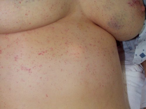
Delayed diagnoses / missed injuries are with us to stay. The typical trauma activation is a fast-paced process, with lots of things going on at once. Trauma professionals are very good about doing a thorough exam and selecting pertinent diagnostic tests to seek out the obvious and not so obvious injuries.

But we will always miss a few. The incidence varies from 1% to about 40%, depending on who your read. Most of the time, they are subtle and have little clinical impact. But some are not so subtle, and some of the rare ones can be life-threatening.

The trauma tertiary survey has been around for at least 30 years, and is executed a little differently everywhere you go. But the concept is the same. Do another exam and check all the diagnostic tests after 24 to 48 hours to make sure you are not missing the obvious.

Does it actually work? There have been a few studies over the years that have tried to find the answer. A paper was published that used meta-analysis to figure this out. The authors defined two types of missed injury:

  • Type I – an injury that was missed during the initial evaluation but was detected by the tertiary survey.
  • Type II – an injury missed by both the initial exam and the tertiary survey

Here are the factoids:

  • Only 10 observational studies were identified, and only 3 were suitable for meta-analysis
  • The average Type I missed injury rate was 4.3%. The number tended to be lower in large studies and higher in small studies.
  • Only 1 study looked at the Type II missed injury rate – 1.5%
  • Three studies looked at the change in missed injury rates before and after implementation of a tertiary survey process. Type I increased from 3% to 7%, and Type II decreased from 2.4% to 1.5%, both highly significant.
  • 10% to 30% of missed injuries were significant enough to require operative management

Bottom line: In the complex dance of a trauma activation, injuries will be missed. The good news is that the tertiary survey does work at picking up many, but not all, of the “occult” injuries. And with proper attention to your patient, nearly all will be found by the time of discharge. Develop your process, adopt a form, and crush missed injuries!

Reference: The effect of tertiary surveys on missed injuries in trauma: a systematic review. Scand J Trauma Resusc Emerg Med 20:77, 2012.

Print Friendly, PDF & Email

The Tertiary Survey for Trauma

Major trauma victims are evaluated by a team to rapidly identify life and limb threatening injuries. This is accomplished during the primary and secondary surveys done in the ED. The ATLS course states that it is more important for the team to identify that the patient has a problem (e.g. significant abdominal pain) than the exact diagnosis (spleen laceration). However, once the patient is ready for admission to the trauma center, it is desirable to know all the diagnoses.

This is harder than it sounds. Physical examination tends to direct diagnostic testing, and some patients may not be feeling pain, or be awake enough to complain of it. Injuries that are painful enough may distract the patient’s attention away from other significant injuries. Overall, somewhere between 7-13% of patients have injuries that are missed during the initial evaluation.

A well-designed tertiary survey helps identify these occult injuries before they are truly “missed.” This survey consists of a structured and comprehensive re-examination that takes place within 48-72 hours, and includes a review of every diagnostic study performed. Ideally, it should be carried out by two people: one familiar with the patient, and the other not. It is desirable that the examiners have some experience with trauma (sorry, medical students).

The patients at highest risk for a missed injury are those with severe injuries (ISS>15) and/or impaired mental status (GCS<15). These patients are more likely to be unable to participate in their exam, so a few injuries may still go undetected despite a good exam.

I recommend that any patient who triggers a trauma team activation should receive a tertiary survey. Those who have an ISS>15 should also undergo the survey. Good documentation is essential, so an easy to use form should be used. Click here to get a copy of our original paper form. We have changed over to an electronic record, and have created a dot phrase template, which you can download here.

Next post: Does the tertiary survey actually work?

Print Friendly, PDF & Email

Clinical Manifestations Of Fat Embolism Syndrome

There are three organ systems that are classically involved in FES: pulmonary, CNS, and skin. Manifestations generally begin between 24 and 72 hours after injury. In rare cases, symptoms can begin within 12 hours. In my experience, these tend to be the ones that become the most severe and are frequently life-threatening.

Pulmonary (95% of cases): This is the most common manifestation of FES, and may occur without other signs and symptoms. Nearly all patients develop some degree of hypoxia. Progressive tachypnea and mild tachycardia may provide the first clinical clue if oxygen saturation is not being monitored.

Chest x-ray is usually unremarkable early on. And once the syndrome has developed, it is generally not helpful. CT scan is useful for defining the extent of pulmonary injury, but lags the clinical picture by several days. Findings are non-specific, usually consisting of small, ground-glass opacities in the periphery.

In the example above, the opacities are very small and difficult to see.

But they’re a little more obvious here!

Other CT findings include small pulmonary nodules in the upper lobes or along peripheral pulmonary vessels. These are thought to be areas of obstruction caused by the emboli. Nonspecific pleural effusions may be seen, and bronchial thickening has also been described. Rarely, fat globules may be seen in the lower extremity veins or IVC, and should immediately raise suspicion for developing FES even before symptoms develop.

CNS (60% of cases): If they occur, CNS changes generally crop up after the pulmonary manifestations begin. Generally, they start as mild confusion, but can progress to decreasing level of consciousness and even coma. Focal neurologic deficits are occasionally seen, and seizures can occur.

The actual mechanism behind this appears to be very similar to the skin changes which will be described in the next section. Emboli occur in vessels predominantly in the white matter of the brain. This leads to petechial hemorrhages, which are likely due to the inflammatory mechanisms previously described.

Note the numerous dark petechiae visible in the white matter in this specimen.

Retinal exam can also show evidence of fat embolism. Fat globules may actually be seen in the retinal vessels early.

Note the fat globules at the 9:30 and 2:00 positions to the optic nerve in the image above.

Skin (33% of cases): The most recognizable sign of FES is the petechial skin rash. This rash usually involves the torso, and axillary petechiae are very common. It can spread to involve the head and neck, and occasionally the extremities. Subconjunctival hemorrhages are sometimes seen. The rash tends to be transient and usually lasts only a few days. Here is an example of the classic petechial rash.

Other findings: Fat globules may be found in the urine in patients with FES. However, they are commonly present in patients with long bone fractures, so their presence is not helpful or predictive. Nonspecific findings such as fever, leukocytosis, anemia, and thrombocytosis are also relatively common. In severe cases, cardiac dysfunction, hypotension, and peripheral hypoperfusion can occur. I have personally seen necrosis of fingers and toes from a very severe case.

Unfortunately, the “classic” triad of mental status changes, skin rash, and pulmonary insufficiency are seen in only a small minority of patients. Typically, only one or two signs and symptoms appear at the same time, making diagnosis a bit challenging.

In the next post, making the diagnosis of fat embolism syndrome.

Print Friendly, PDF & Email

Animals vs Cars: What To Do

This is a bad time of year in much of the United States for striking animals on the road. In my part of the country, the deer are out in full force. Car vs animal can be challenging, and motorcycle vs animal is frequently deadly. What can our patients do to protect themselves?

  • Be especially vigilant when driving for the first few hours after sunset and just before sunrise. More animal activity occurs during these hours.
  • If one animal is spotted, look out for others.
  • Drive with high beams on as much as possible. In many animals, this will show reflections from their eyes. Some large animals, such as moose, don’t have glowing eyes.
  • Always where a seat belt in case an impact does occur.
  • If an animal is spotted, slow down quickly and blow the horn.

Most important! NEVER swerve or attempt to quickly change direction. This is one of the most common errors that results in serious injury or death. The driver swerves to avoid the animal and begins to leave the roadway. They then over-correct in the opposite direction, triggering a rollover. Always make gentle corrections, staying in the same lane.

For small animals, slowly adjust the steering wheel to straddle them with the wheels. For larger ones, try to plan the impact so it is in front of the unoccupied front passenger seat. If the seat is occupied, plan the strike in the middle of the hood. The idea is to keep the car occupants safe, but to assist with natural selection and remove the animal from the gene pool.

Print Friendly, PDF & Email

Best Of AAST 2021: Trauma Transfers Discharged From The ED

Aren’t these embarrassing? A referring center sends you a patient with the idea that they will be evaluated and admitted to your hospital. But it doesn’t work out that way. The patient is seen, possibly by a surgical specialist, bandaged up, and then sent home. Probably to one that is quite a few miles away. Not only is this a nuisance for the patient and an embarrassment for the sending center, it may use resources at the trauma center that are already tight.

Transfer patients who are seen and discharged are another form of “ultimate overtriage.” In this case, the incorrect triage takes place at the outside hospital.  The trauma group in Oklahoma City reviewed their experience with these patients over a two year period. They looked exclusively at patients who were transferred in to a Level I center and then discharged.

Here are the factoids:

  • A total of 2,350 patients were transferred in, and 27% were transferred home directly from the trauma bay (!)
  • The three most common culprits by injury pattern were face (51%), hand (31%), isolated ortho injury (9%)
  • A third of these patients required a bedside procedure, including laceration repair (53%), eye exam (24%), splinting (18%), and joint reduction (5%)
  • Ten facilities accounted for 40% of the transfers

The authors concluded that the typical injuries prompting transfer are predictable. It may be possible to reduce the number of transfers by deploying telemedicine systems to push evaluations out to the referring hospitals.

Bottom line: This is quite interesting. Anyone who works in a Level I or II center is aware of this phenomenon. This abstract went a step further and quantified the specific issues involved. This center ended up discharging over 300 patients per year after transfer in. This is a tremendous drain on resources by patients who did not truly have the need for them.

The authors speculate that telemedicine evaluation may help reduce some of those transfers. This seems like an easy solution. However, it also poses a lot of issues in terms of who will actually staff the calls and how will they be compensated for their time.

There are a number of important take-aways from this abstract:

  1. Know your referring hospitals. In this study, there were 10 hospitals that generated an oversize number of referrals. Those are the targets / low hanging fruit. Identify them!
  2. Understand what their needs are. Are they frequently having issues with simple ortho injuries? Eye exams? This is what they need!
  3. Provide education and training to make them more comfortable. This allows you to target those hospitals with exactly the material they need and hopefully make them more self-sufficient.

This allows the higher level centers to reserve phone and/or telemedicine consultation for only the most ambiguous cases. It’s a better use of telehealth resources that may be needed, typically at night and on weekends.

Here are my questions for the presenter and authors:

  • Would the common issues that were transferred and discharged be amenable to education and training at the referring centers to decrease the transfer volume?
  • How have you begun to address this issue at your center?

Reference: TRAUMA TRANSFERS DISCHARGED FROM THE EMERGENCY DEPARTMENT – IS THERE A ROLE FOR TELEMEDICINE. AAST 2021, Oral abstract #63.

Print Friendly, PDF & Email