Category Archives: General

Who Travels By Air?

Getting seriously injured trauma patients to a trauma center quickly is generally believed to be a good thing. And helicopters are usually faster than ground ambulances. So sending severely injured patients by air is a good thing, right?

Not quite so fast, there. There are other concerns as well. Helicopter transport is significantly more expensive. Quarters are very cramped, and you can’t just pull off to the side of the road if major patient or equipment problems arise. And has anybody really shown a survival benefit?

Although there is a (relatively) standard national trauma triage protocol from the CDC that indicates which patients should be transported to a trauma center, there is no standard protocol for who should be transported by air. The University of Rochester School of Medicine looked at 2007 transport data from the National Trauma Databank and tried to determine if the CDC protocol could be adapted to air transport as well.

Over 250,000 patient records were included in the study. As would be expected, patients flown by helicopter tended to be more severely injured, needed intubation more often, and were admitted to an ICU and stayed in the hospital longer. Average transport time for the helicopter was longer (60 mins vs 43 mins), implying longer distance traveled. Using a regression analysis, the authors found that the following subsets of patients had better survival with helicopter transport:

  • Penetrating injury
  • GCS < 14
  • Resp rate <10 or >29
  • Age >55
  • Any one physiologic criterion and any one anatomic criterion from the CDC protocol

Bottom line: A more standardized set of air transport criteria is needed. Some studies have found that as many as 50% of patients in some communities are flown who do not meet local air transport rules. Time and distance also need to be taken into account, since these will vary widely between rural and less rural areas. This study begins to lay an objective framework of criteria that can be incorporated into a more uniform set of guidelines.

Related posts:

Reference: The National Trauma Triage Protocol: Can this tool predict which patients with trauma will benefit from helicopter transport? J Trauma 73(2):319–325, 2012.

Pop Quiz! DPL – The Answer!

You’re doing one of those (very rare) DPLs and get a surprise result. Not blood, not obvious intestinal content, but just a small amount of mysterious sediment. What to do?

Well, this is obviously not normal. Therefore, this has to be considered a positive diagnostic peritoneal lavage. Since DPL is a qualitative test (meaning that the answer is only yes or no), the patient must go to the OR.

Here are the answers to the questions posed earlier today:

  • The DPL catheter has a relatively small diameter, so leave it in place! It may be very difficult to find where it went otherwise
  • Midline laparotomy incision is most appropriate. Remember, this is a trauma case? However, you can start infra-umbilical with a limited incision.

Here’s what I found in this case:

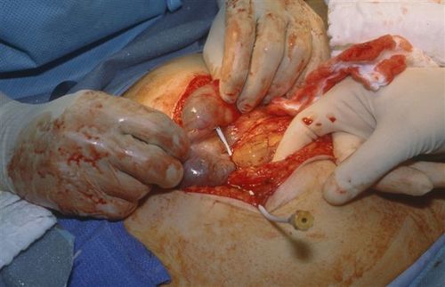
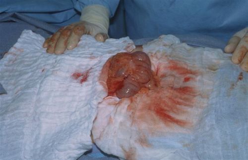
The catheter went straight into the cecum! So we actually did a diagnostic colonic lavage! The sediment was a very small amount of stool. And as stated above, had the catheter not been left in place, it would have been very tough to find the puncture site. 

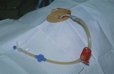
Next, I clamped the catheter to keep it in place, cut it on the hub side, and removed most of it.

Finally, I placed a purse-string stitch around the entry site in the bowel, removed the catheter and tied the suture.

But wait, we’re not done yet! The patient did have abdominal pain and a seat belt sign, so we did a trauma exploration through the midline incision. A Grade II liver injury was present which needed no further management. The patient did well  and was discharged on the fourth day.

Bottom line: Procedures can and do go awry. Reason your way through it the best you can, then use focused diagnostics, if needed, to come up with a plan. For misplaced needles and catheters, most organs can tolerate a puncture by almost anything (except the eye, maybe). Treat appropriately and monitor carefully afterwards.

Source: Personal archive. Not treated at Regions Hospital

Pop Quiz! DPL Hint

So the catheter is in, the aspirate was negative (nothing came out), and a liter of crystalloid infused easily. But toward the end of draining the fluid back out, some faint sediment became visible in the tubing.

A lot of you guessed bladder, but most people don’t have sediment there. Plus, if I dumped a liter of fluid into your bladder, you’d really get the urge to go. This awake patient noted no new symptoms. 

I had a bad feeling about this, so I elected to take her to the OR to see what the story really was. Here are some questions for any budding surgeons out there:

  • Leave the catheter in place or pull it out before OR?
  • What incision to make?
  • How big?
  • And what the heck is it, really?

Answers later today! See if you can get it before I give you the punch line!

Pop Quiz! DPL

Ahh, remember the good old days of DPL? Probably not! But here’s an interesting case that presents a real diagnostic dilemma. Hint: this case occurred B.F. (before FAST) and B.G.C.T. (before good CT). That’s why we used DPL!

The patient was a middle aged woman who was involved in a car crash. She had mild, diffuse abdominal pain and a faint seat belt sign. She was prepared for DPL in the ED. It was performed using percutaneous (Seldinger) technique with a fenestrated catheter. Placement was in the usual position, 2cm below the umbilicus in the midline.

The aspirate was negative. A liter of LR was infused  and the bag was then lowered to drain. About 600 cc of clear amber fluid returned easily.

However, on closer inspection, a small amount of sediment could be seen in the tubing.

What the heck!? What’s going on and what, if anything, do we need to do?

Post your guesses and comments below, or Tweet them. I’ll provide hints over the weekend, and the answer on Monday.

Source: Personal archive. Not treated at Regions Hospital

Fracture Blisters Demystified

Fracture blisters pop up (!) in trauma patients now and then, and nobody seems to know what to do with them. Here’s a primer on dealing with them.

A fracture blister typically occurs near fractures where the skin has little subcutaneous tissue between it and bone. These include elbows, knees, ankles and wrists. They tend to complicate fracture management because they interfere with splinting, casting, and incision planning for open reduction procedures. They can appear anytime within a few hours of injury to 2-3 weeks later.

These blisters are thought to be caused by shearing forces applied at the time of injury. There are two types described, based on their color: clear fluid and hemorrhagic. The difference lies in the level of the shear. Clear fluid blisters have separated within the epidermis, and hemorrhagic blisters separate at the dermal-epidermal junction. The clinical difference is healing time; clear blisters take about 12 days and hemorrhagic blisters heal in about 16 days. 

So should we pop the blisters and operate/splint, or wait for them to heal and then go to surgery? Unfortunately, there’s no great data on this and it usually hinges on the preferences of the orthopaedic surgeon. Waiting delays care an average of 7 days, and longer for tibial plateau and calcaneal fractures. Operating immediately anecdotally increases wound infection rates.

Bottom line: Anticipate fracture blisters by looking at location and severity of mechanism. Try to schedule operative reduction as soon as is practical. And monitor the wound closely to make sure that delayed blisters don’t cause complications due to splinting or casting.