Category Archives: General

Antibiotics For Ventriculostomy

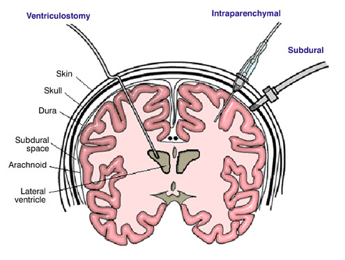
There is some variability in how neurosurgeons manage ventriculostomy when it comes to antibiotic coverage. The catheter can become a conduit for bacterial infections of the meninges or brain, which can be life threatening. Most neurosurgeons will begin coverage at the time of catheter insertion, but the duration of treatment varies. What is the right answer, if any?

As is typical with most things related to head trauma, there are not a lot of good studies out there. Columbia University published a fairly comprehensive review of previous studies last year to help clarify this issue. They applied rigorous criteria to identify 10 relevant studies (3 randomized clinical trials and 7 observational studies) out of a pool of 347. Yes folks, this gives you an idea of how tough it is to answer good clinical questions from the stuff that gets published.

The study found that the use of either prophylactic antibiotics or antibiotic-coated external ventricular drains (EVD) decreased the number of infections by 68%. This result was consistent across both study designs. The authors could not show that one mode of antibiotic administration (systemic vs catheter coating) was better than the other. About half of the studies used antibiotics for the duration of the catheter; the other half did not specify.

Bottom line: Head injury patients with an EVD should receive antibiotics, either systemically or as a coating on the EVD catheter itself. Although not entirely clear, they should probably be given for the entire time the catheter is in place. Judgment must be used if this will be a long time, because there may be other adverse effects from giving long term antibiotics.

Reference: Prevention of Ventriculostomy-Related Infections With Prophylactic Antibiotics and Antibiotic-Coated External Ventricular Drains: A Systematic Review. Neurosurgery 68(4):996-1005, 2011.

Print Friendly, PDF & Email

Air Travel After Pneumocephalus: Part 2

I’ve previously written about air travel after pneumothorax. The issue is that air expands as you ascend to aircraft level heights. That means that air bubbles in your chest can double or almost triple in size on the average airliner at altitude. For the official recommendations for when it is safe to travel after a pneumothorax, see the link to my previous post below.

And I covered pneumocephalus previously as well. This is a very infrequent condition, and there is very little literature about it. After doing a thorough search, I found two additional papers on it. The first was a computer simulation from Sweden. As expected, it found that air bubbles expand 30% when cabin altitude reaches 8000 ft. They were also able to model changes in intracranial pressure (ICP), and found that it increased as well. The amount was related to the size of the original air bubbles and the rate of the ascent (faster ascent = higher ICP). The worst case ICP increase with a 30cc bubble was 11 torr.

The other paper was from the UK and described a survey of neurosurgeons regarding their advice to patients on air travel after craniotomy. The fear is that there may be residual bubbles around the brain from the surgery. The results of this type of survey are usually all over the place, and more so when you ask neurosurgeons. To their credit, 92% advised their patients not to fly for a period of time. Unfortunately, the timeframes ranged from <2 to >8 weeks!

How about some practical info? I previously wrote about an actual clinical study by the US military who transported soldiers with pneumocephalus by air. Their series of 21 patients showed no neurological deterioration during flight, and 3 who had ICP monitors had no significant changes in pressure.

Bottom line: Computer models (and common sense) tell us that it’s a bad idea to fly commercially with air around your brain. It may work for the military because they have to fly, and the soldier is in a flying ICU. But in civilian aircraft, there is little to be done if medical problems occur short of an emergency landing and transport to a hospital. What’s the magic number of weeks to wait to fly? That is not known, but 2 weeks has worked well for pneumothorax so it seems like a reasonable place to start. But it should be easy for someone to do a retrospective review of serial head CTs from craniotomy patients to see how long it takes for the bubbles to actually reabsorb. And don’t worry about helicopter flights; altitudes are too low to make a difference!

Related post:

References:

  • Air transport of patients with intracranial air: computer model of pressure effects. Aviat Space Environ Med Feb 74(2):138-44, 2003.
  • Air travel after intracranial surgery: a survey of advice given to patients by consultant neurosurgeons in the UK. Br J Neurosurg 2012 Aug 30, epub ahead of print.
Print Friendly, PDF & Email

TraumaMedEd Newsletter for August

The newest TraumaMedEd newsletter is out! It was emailed to subscribers on Monday. This months focus is prehospital, with articles on the following:

  • Hare traction application
  • Trauma care and HIPAA, demystified
  • Trauma patient survival and air transport
  • What happens when you violate resuscitation guidelines for traumatic cardiac arrest?
  • Handoff issues between prehospital and the trauma team

Download the newsletter here!

Subscribe to the newsletter by clicking here!

Print Friendly, PDF & Email

Trauma Residents: How To Remember Liver Anatomy

In trauma surgery, operative management of liver injury is usually messy business, with little time for nice anatomic resections. However, an understanding of the basic anatomy, especially that of the vascular supply is crucial for saving your patient.

A cool tool for remembering Couinaud’s segments and the overall layout of liver anatomy was published in the Archives of Surgery recently. It makes use of a model, which consists of your hand! Just make a fist with your right hand and tuck the thumb behind the other fingers.


The fingers can then be numbered according to the Couinaud segments, with the caudate lobe (segment 1) represented by the thumb that is tucked away. The PIP joints represent the plane that the portal vein runs through, with branches going to upper and lower segments. Note how the ring finger normally lies a little more anterior than the little finger in this position, just like the sectors of the right lobe.

The creases between the fingers represent the left, middle and right hepatic veins.


The right hepatic vein is located between the right anterior and posterior sectors and the left hepatic vein sits between the left medial and lateral sectors. The middle hepatic vein is in between the left and right hemi-liver.

Bottom line: This “handy” liver model is available immediately in the OR and is already sterile. It can help visualize liver structures that may be injured quickly and accurately to speed your operative approach to the problem.

Reference: A Handy Tool to Teach Segmental Liver Anatomy to Surgical Trainees. Arch Surg 147(8):692-693, 2012.

Print Friendly, PDF & Email