Category Archives: Extremity

Giving Vitamin D After Fracture

The role of Vitamin D in fracture healing is well known. So, of course, trauma professionals have tried to promote Vitamin D

supplementation to counteract the effects of osteoporosis. A meta-analysis of of 12 papers on the topic relating to hip and other non-vertebral fractures showed that there was roughly a 25% risk reduction for any non-vertebral fractures in patients taking 700-800 U of Vitamin D supplements daily.

Sounds good. So what about taking Vitamin D after a fracture occurs? Seems like it should promote healing, right? A very recent meta-analysis that is awaiting publication looked at this very question.

Unfortunately, there was a tremendous variability in the interventions, outcomes, and measures of variance. All the authors could do was summarize individual papers, and a true meta-analysis could not be performed.

Here are the factoids:

  •  81 papers made the cut for final review
  • A whopping 70% of the population with fractures had low Vitamin D levels
  • Vitamin D supplementation in hospital and after discharge did increase serum levels
  • Only one study, a meeting abstract which has still not seen the light of day in a journal, suggested a trend toward less malunions following a single loading dose of Vitamin D

Bottom line: Vitamin D is a great idea for people who are known to have, or are at risk for, osteoporosis and fractures. It definitely toughens up the bones and lowers the risk of fracture. However, the utility of giving it after a fall has not been shown. Of the 81 papers reviewed, none showed a significant impact on fracture healing. The only good thing is that Vitamin D supplements are cheap. Giving them may make us think that we are helping our patient heal, but it’s not. 

References:  

  • What is the role of vitamin D supplementation in acute fracture patients? A systematic review and meta-analysis of the prevalence of hypovitaminosis D and supplementation efficacy. J Orthopaedic Trauma epub Sep 22 2015.
  • Fracture prevention with vitamin D supplementation: a meta-analysis of randomized controlled trials. JAMA 293(18):2257-2264, 2005.

Best of AAST #9: Popliteal Atery Injury Repair

Injury of the popliteal artery is potentially devastating. Since this vessel is essentially and end artery, any complication resulting in thrombosis can result in limb loss. Traditionally, significant injuries have been treated with open repair and/or bypass. However, endovascular therapies have been making inroads in this area. Short-term outcomes appear to be equivalent. But what happens in the long term? Is one better than the other?

Scripps Mercy in San Diego (yes, same as yesterday’s abstract!) performed a retrospective review of the same California state discharge database. This time, they focused on patients with popliteal artery injury, and the attendant complications of fasciotomy and amputation. They stratified the patients into open and endovascular groups.

Here are the factoids:

  • 769 patients with popliteal artery injury were identified over an 8-year period
  • 59% were managed with an open operation, 4% using endovascular techniques, 2% combined, and 34% nonoperatively
  • Fasciotomies were performed significantly more often in the open group (41% vs 19%)
  • More amputations were performed in open cases, but this was not significantly different (11% vs 3% [1 patient in the endovascular group])
  • Embolism or thrombosis was significantly more likely during the first admission in endovascular or combined endo/open cases
  • Patients requiring both endo and endo+open procedures  were 5x more likely to undergo a later amputation, and 4x more likely to die after discharge

Bottom line: First, remember the limitations of this study: (very) small numbers, and a large database that precludes teasing out details. It suggests that open repair of popliteal injury is superior to endovascular due to higher thrombosis/embolism and amputation rates. Performing a fasciotomy is somewhat subjective, and may be done by surgeon preference to protect the limb. But amputation is more objective.

Unfortunately, we will not get anything more definitive any time soon. This 8-year analysis of a huge state database yielded only 769 cases, or 96 per year. In a state with 39 million people. That’s three injuries (reported) per million people per year. We will never generate a study that will tell us the full answers. But in the meantime, consider endovascular repair of popliteal artery injury only in patients for whom an open procedure is more challenging or risky (e.g. obesity, associated wounds).

Reference: Outcomes for popliteal artery injury repair after discharge: a large-scale population-based analysis. Session XXII Paper 55, AAST 2018.

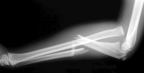
What Is: The Monteggia Fracture

Yesterday, I wrote about one of the many fractures that occurs during falls onto outstretched hands, the Galeazzi fracture. Today, I’ll describe another one, the Monteggia fracture. Yes, this one is named after another Italian surgeon! And like the other one, the person it was named after was actually the second to describe it.

Think of the Monteggia fracture as the exact opposite of a Galeazzi fracture. The fractured bone is switched, as is the dislocation. Whereas the Galeazzi is a distal radius fracture with a distal ulnar dislocation which pulls the radio-ulnar joint apart, the Monteggia is a proximal ulnar fracture with a proximal radial head dislocation.

Here’s what it looks like:

Of course, the orthopedic surgeons have a classification system for this based on the directions the bones fracture and dislocate. I won’t bore you with the details.

Unlike the Galeazzi fracture, all of these require operative repair, even in children. This helps stabilize the radial head and decreases the incidence of malunion.

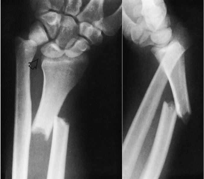
What Is: The Galeazzi Fracture?

Orthopedic surgeons have so many names for fractures, it gets confusing! Today, let’s dig in to the “Galeazzi fracture.” This one was named for an Italian surgeon during the early 20th century) although it was actually first described by an Englishman named Cooper a hundred years earlier).

The Galeazzi fracture is an uncommon one, and consists of two components: a radius fracture at the junction of the distal and middle thirds, and a dislocation of the distal radio-ulnar joint. Here’s what it looks like:

Notice the obvious dislocation seen in the lateral view. Of course, a whole classification system has been developed to describe the various nuances of this fracture pattern, but that’s beyond the scope of this post.

What to do about it? This one needs prompt orthopedic consultation, and due to the dislocation component it requires operative management in adults. In children, initial closed reduction is the treatment of choice.

Monday, I’ll describe this fracture’s evil twin, The Monteggia fracture.

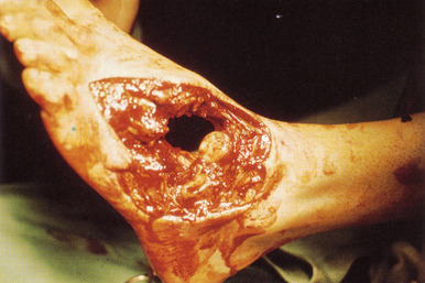
What A MESS! Part 2

The trauma season is always officially open. And unfortunately, our patients can sustain mangled extremities in any number of ways. In days of old, management was simple: take it off. But we’ve become wiser over the years and are now able to salvage a good number of these threatened limbs. The Mangled Extremity Severity Score (MESS) has helped greatly with this. 

As I mentioned yesterday, it’s beginnings were humble, almost looking like guesswork on the part of the authors. But this system has withstood the test of time.

There are four components to MESS: limb ischemia, patient age, presence of shock, and mechanism of injury. Each component is assigned an integer value depending on severity. The possible values range from 1 to 14. Here’s the breakdown of each component:

Ischemia

  • +1 Reduced pulse but normal perfusion
  • +2 Pulseless, paresthetic, reduced capillary refill
  • +3 Cool, paralyzed, insensate
  • Add 3 points if limb ischemia has been present more than 6 hours

Age

  • +0   <30 years
  • +1   30-50 years
  • +2   >50 years

Shock

  • +0 SBP >90 consistently
  • +1 Transient hypotension
  • +2 Persistent hypotension

Mechanism (kinetic energy)

  • +1 Low (stab, gunshot, simple fracture)
  • +2 Medium (dislocation, open or multiple fractures)
  • +3 High (high speed MVC, rifle)
  • +4 Very high (high energy trauma with gross contamination)

Per the original study, values of 7 or greater predict low salvageability. However, with advancing technology, drugs, and operative techniques, the threshold has been creeping higher. But not that much higher, probably 8 or so.

Bottom line: Use the MESS score as one tool in your armamentarium to help address mangled extremities. But remember, it is not the final answer. In the OR, confer with your orthopedic and vascular colleagues. Decide if immediate amputation is necessary, or whether a second look in a day or two is in order. Use MESS as a tiebreaker. But remember, don’t let your desire to save the extremity jeopardize your patient’s life (rhabdomyolysis, renal failure, acidosis). If systemic signs begin to occur, cut your (and their) losses and amputate!

Related post: