Category Archives: Extremity

Vascular And Nerve Injury After Knee Dislocation

There’s a lot of dogma in trauma care, as well as in the field of medicine generally. The knee dislocation dogma is that the incidence of vascular injury is high (around 50%) with posterior dislocation and somewhat lower with non-posterior dislocation.

At least, that’s what I learned way back when. After recently finding myself spouting off those numbers, I wondered if it was really true. Our diagnostic imaging and vascular care have increased considerably in the last few decades, so I decided to check it out.

This lovely image from EMDocs.net shows the various dislocation types. It also gives you an idea of why an associated vascular or nervous injury is so common.
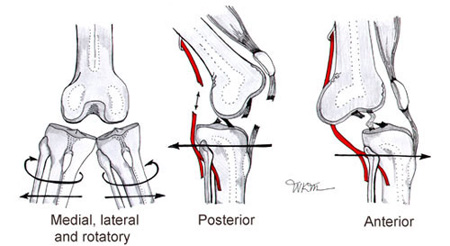
(The nomenclature of the dislocation is based on the direction in which the tibia and fibula move with respect to the femur.)

The orthopedic surgery group at UCLA performed a meta-analysis of the literature on knee dislocation complications. They identified seven papers describing the injuries of 862 patients.

Here are the factoids:

  • The overall incidence of vascular injury with knee dislocation was 18%, and nerve injury was 25%
  • The incidence of vascular injury with the various types of dislocation was:
    • Posterior dislocation: 25%
    • Anterior dislocation: 19%
    • Lateral dislocation: 18%
    • Medial dislocation: 7%
    • Rotatory: 14%
  • Disruption of both cruciate ligaments as well as the lateral or medial collateral ligament had a very high incidence of vascular injury (32% and 26% respectively)
  • About 80% underwent surgical repair of the popliteal artery, but the amputation rate was 12%(!)

Bottom line: The old dogma regarding vascular injury after knee dislocation may be slightly exaggerated. However, it is still common after knee dislocation and can lead to devastating complications.

If your patient tells you that they felt a popping sensation in their knee or have a mechanism consistent with knee dislocation (e.g., pedestrian struck), be very suspicious for this injury. A thorough yet gentle exam, including good neurologic and vascular exams, should be performed. Calculating the Arterial Pressure Index (API) may be helpful but will not keep you from obtaining imaging studies. Multi-plane knee imaging is required, and a CT angiogram/runoff study should be performed to exclude a vascular problem.

Reference: Vascular and Nerve Injury After Knee Dislocation: A Systematic Review. Clin Orthop Relat Res 472(9):2621-9, 2014.

Best of AAST 2023 #2: Immediate Postoperative Prosthesis

Blunt vehicular trauma is the most common cause of severe lower extremity trauma, particularly motorcycle crashes. Occasionally, the injury is so severe that the limb cannot be saved, and amputation is necessary. The conventional treatment is to protect the amputation incision, provide physical therapy, and fit a prosthesis once the stump is mature. This typically takes a month or two.

Unfortunately, losing any limb has a significant psychological impact on our patients’ physical and mental well-being. The concept of immediate postoperative prosthesis (IPOP) has been gaining traction in recent years in an attempt to improve early mobility and mental health among these amputees.

A group from India designed a randomized, controlled trial to compare patients undergoing IPOP after lower extremity amputation to those receiving conventional prosthetic treatment. They randomly enrolled 30 patients in each group and measured differences in quality of life, depression and anxiety, and various mobility scores.

Here are the factoids:

  • Both groups were modestly injured, with 85% having ISS < 15; this indicates that injuries were mostly limited to the extremity
  • Mangle extremity severity score was also low, indicating the incidence of vascular and severe soft tissue injury was also low
  • Quality of life scores for the physical, psychological, social, and environmental domains were significantly higher in the IPOP group
  • The Amputee Mobility Predictor score (AMP) was significantly higher after 12 weeks after IPOP
  • The Trinity Amputee Prosthesis Experiences Scales (TAPES) for psychosocial, activity restriction, and prosthetic satisfaction domains were significantly better in the IPOP group
  • The physical screening tests for directional control and overall stability were also significantly higher in IPOP patients

The authors concluded that IPOP improves quality of life, decreases depression and anxiety, and increases mobility in amputees compared to standard therapy.

Bottom line: It is common sense that allowing early mobility would help our patients, both physically and mentally. This paper makes it clear that IPOP makes a very real difference. This small study bears additional confirmatory work, but given the level of significance found, the concept will likely be proven.

It does take some extra effort to apply a well-fitted early prosthesis. This typically takes place in the OR. The prosthesis must be easy to remove for wound care and protect the stump from injury while weight-bearing.  It is best done by an orthopedic surgeon and skilled prosthetist at the end of the amputation procedure. 

Hopefully, this concept will catch on to help patients with this potentially devastating procedure recover more quickly and retain their mental health.

Reference: RCT to study the effect of immediate post-operative prosthesis vs. conventional prosthesis on balance & QOL in BK amputees following trauma. AAST 2023, Plenary paper #21.

Novel Hip Reduction Technique: The Captain Morgan

I wrote about posterior hip dislocation and how to reduce it using the “standard” technique quite some time ago (see link below). Emergency physicians and orthopedic surgeons at UCSF-Fresno have published their experience with a reduction technique called the Captain Morgan.

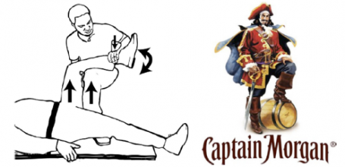
Named after the pose of the trademark pirate for Captain Morgan rum, this technique simplifies the task of pulling the hip back into position. One of the disadvantages of the standard technique is that it takes a fair amount of strength (and patient sedation) to reduce the hip. If the physician is small or the patient is big, the technique may fail.

In the Captain Morgan technique, the patient is left in their usual supine position and the pelvis is fixed to the table using a strap (call your OR to find one). The dislocated hip and the knee are both flexed to 90 degrees. The physician places their foot on the table with their knee behind the patient’s knee. Gentle downward force is placed on the patient’s ankle to keep the knee in flexion, and the physician then pushes down with their own foot, raising their calf. Gentle rotation of the patient’s hip while applying this upward traction behind the patient’s knee usually results in reduction.

Some orthopedic surgeons use a similar technique, but apply downward force on the patient’s ankle, using the leverage across their own knee to develop the reduction force needed. The Captain Morgan technique use the upward lift from their own leg to develop the reduction force. This may be gentler on the patient’s knee.

The authors report a series of 13 reductions, and all but one were successful. The failure occurred due to an intra-articular fragment, and that hip had to be reduced in the operating room.

Reference: The Captain Morgan technique for the reduction of the dislocated hip. Ann Emerg Med 58(6):536-540, 2011.

Posterior Hip Dislocation

Although posterior hip dislocation is an uncommon injury, the consequences of delayed recognition or treatment can be dire. The majority are caused by head-on car crashes, and 90% of these are posterior dislocations. The femoral head is forced across the back wall of the acetabulum, either by the knee striking the dash, or by forces moving up the leg when the knee is locked. This occurs most commonly on the right side when the driver is standing on the brake pedal, desperately trying to stop.

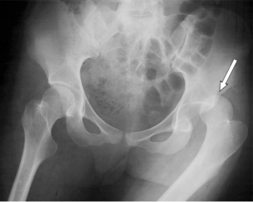
On exam, the patient presents with the hip flexed, internally rotated and somewhat adducted. Range of motion is limited, and increasing resistance is felt when you try to move it out of position. An AP pelvic X-ray will show the femoral head out of the socket, but it may take a lateral or Judet view to tell if it is posterior vs anterior.

These injuries need to be reduced as soon as possible to decrease the chance of avascular necrosis of the femoral head. Procedural sedation is required for all reductions, since it makes the patient much more comfortable and reduces muscle tone. The ED cart needs to be able to handle both the patient’s weight and your own. I also recommend a spotter on each side of the cart.

Standing on the cart near the patient’s feet, begin to apply traction to the femur and slowly flex the hip to about 90 degrees. Then gently adduct the thigh to help jump the femoral head over the acetabular rim. You will feel a satisfying clunk as the head drops into place. Straighten the leg and keep it adducted. If you are unsuccessful after two tries, there is probably a bony fragment keeping the head out of the socket. See an instructional video on this in my next post.

Regardless of success, consult your orthopedic surgeon for further instructions. And be sure to thoroughly evaluate the rest of the patient. It takes a lot of energy to cause this injury, and it is flowing through the rest of the patient, breaking other things as well.