Category Archives: Equipment

Why Use A Hybrid OR For Trauma?

Trauma is a surgical disease, and specifically, a disease of bleeding. So many of the tools and processes we have developed for its management revolves around the control of hemorrhage.

When a major trauma patient arrives in the resuscitation room, the initial management involves rapid assessment and correction of life-threatening conditions. Recognition of bleeding is paramount. A rapid decision must be made as to the source of hemorrhage and the best way to control it.

Traditionally, bleeding control has been relegated to the operating room. Body cavities are opened as appropriate, and exsanguination is controlled by clamping, repairing, and/or suturing.

However, some body regions are much more challenging. The most notable is the pelvis, and specifically, the unstable pelvis. In the old days, after wrapping or applying an external fixator, the best we could do was to ligate the internal iliac arteries bilaterally and hope the bleeding would slow down sufficiently (it never really stopped) so that internal packing might have a chance.

As the use of interventional radiography grew in trauma, it became possible to noninvasively occlude the internal iliacs. And then, the radiologists became skilled enough that they could selectively identify and embolize more distal bleeding vessels that would dramatically shut down pelvic bleeding.

But this introduced a conundrum. OR vs IR? Where to go after the trauma bay? I’ve long said that the only place an unstable trauma patient can go is to the OR. Not CT, and certainly not the radiology department.

Only the OR, because that’s the only place that something can actually be done about the bleeding. However, that’s not entirely true now.

Here’s the traditional algorithm for a patient with hemorrhage from pelvic fractures:

They go to the operating room OR interventional radiology. If they start in the operating room and can be stabilized (think external fixation and/or preperitoneal packing), then they might be able to be packaged and taken to IR for embolization. And likewise, if they were initially stable enough to go to IR but crash there, then they must immediately be taken to OR.

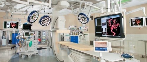
But what if you could do both in one room?! That’s the beauty of the hybrid room! It is entirely possible to do two, three, and maybe more cases on the same patient in the same room. Hence, the hybrid OR.

Tomorrow, is the hybrid OR for trauma useful?

What Is A Hybrid OR, Exactly?

A hybrid operating room is a special suite that allows advanced imaging to be carried out at the same time as one or more additional operative procedures. It’s that simple. It contains specialized imaging equipment including fluoroscopy and infusion equipment for radiographic dye administration. Some also contain CT and/or MRI capabilities, although the shielding required for these makes them very rare. It is generally stocked with a variety of endovascular

devices and supplies. The usual anesthesia circuits are available, as are selected surgical packs, typically related to vascular and CV surgery.

These suites are typically large, and can easily accommodate multiple operating teams. However, they are very expensive in a number of ways.

First, they take up a great deal of space. Many have the square footage of two or more standard operating rooms. Initial construction costs are very high, as are remodeling and maintenance costs. They can also tax the hospital engineering infrastructure, from electrical to plumbing to ventilation.

But if a hybrid room is available, it can deliver significant benefits to the hospital and to patient care. Intraoperative imaging can provide immediate quality assurance, and patients can undergo more complex procedures and enjoy a shorter length of stay.

Tomorrow, why use a hybrid room for trauma?

It’s Hybrid OR Week!

While quite a bit of trauma care is routine, involving simpler, single system injuries, a small subset of our patients sustains major, multi-system, and life-threatening ones. They require rapid access to skilled trauma professionals and advanced resources including imaging, operating rooms, and other procedures.

In most trauma centers, initial resuscitation takes place in a trauma resuscitation room in or near the ED. Some diagnostic imaging can be performed there, but more sophisticated studies may require a short (or longer) road trip. Operating rooms and other procedural areas are also usually more distant. And most importantly, each of these areas is designed for a single discipline. Diagnostic radiology has equipment, technicians, and radiologists available. Interventional radiology contains the specialized equipment needed for this more invasive procedure. ORs are designed specifically for surgical procedures, and frequently contain equipment for a single surgical discipline.

But some of our patients require it all! Think about a patient who arrives after a major car crash. Blood pressures are soft, the pelvis is grossly unstable, FAST exam is positive, and there is bleeding from the vagina.

How do we prioritize? Where do we go first? How long will it take the interventional radiology team to arrive? Where’s that external fixator equipment? Can we slip in a CT scan? Where’s OB/GYN??

The solution is right under our nose! Many hospitals have added so-called “hybrid ORs” to their operating suites in order to address the needs of their vascular and cardiovascular surgeons. This week I’ll cover everything you need to know about this important tool for trauma care. I’ll review:

  • What is a hybrid OR, exactly?
  • Why use a hybrid OR for trauma?
  • Is the hybrid OR for trauma useful?
  • Which patients may benefit from a hybrid OR?
  • So you want your own hybrid room?!

Tomorrow, what is a hybrid OR, exactly?

Trends In IVC Filter Placement And Retrieval

Yesterday, I reviewed a paper that highlighted a single-institution experience for IVC filter usage. Today, let’s look at a much larger pool of data.

Placement of a filter in the inferior vena cava (IVC) is one of the many tools for managing pulmonary embolism. There was a significant increase in filter placement during the 1990s and 2000s due to a broadening of the indications for its use.  There has been continuing debate over the complications and efficacy of use of this device.

A paper from NYU Langone Health in New York City, the Harvey L. Neiman Health Policy Institute, and Georgia Institute of Technology School of Economics looked a long-term trends in IVC filter use in the Medicare population. They scanned a Centers for Medicare and Medicaid Services (CMS) database over the 22 year period from 1994 to 2015. They specifically analyzed trends in insertion, removal, placement setting, and specialty of the inserting physician.

Here are the factoids:

  • 2008 seemed to be the heyday of IVC filter insertion. Rates nearly tripled by 2008, but have declined about 40% since then (see below). Pay attention to the retrieval rates.

  • Overall, filters were most commonly placed by radiologists, followed by surgeons and cardiologists. Here’s the diagram above broken down by specialty.

  • This chart shows the market share of each specialists inserting IVC filters during the study period. Of note, radiologists continue to increase and surgeons are decreasing.

Bottom line: This study shows some interesting data, but can’t be completely applied to trauma patients because it focuses on Medicare recipients. But the trends are valid. IVC filter use peaked in 2008 and has been declining ever since. Radiologists place more filters than other specialties, and their market share continues to increase.

Most disturbing is the low filter retrieval rate, similar to what was seen in yesterday’s post. Device manufacturers recommend removal of most filters, but timeframes are not specified. The real bottom line is that we have an indwelling device which works well in very limited situations only, can cause long term complications, and that we frequently forget to remove. It behooves all trauma professionals to develop strict guidelines for both use and removal.

Reference: National Trends in Inferior Vena Cava Filter Placement and Retrieval Procedures in the Medicare Population Over Two Decades. J Am Coll Radiol 15:1080-1086, 2018.

What’s The Best Pelvic Binder? Part 2

Yesterday, I detailed some pelvic binders commonly available in the US. Today, I’ll go through the (little) science there is regarding which are better than others.

There are a number of factors to consider when choosing one of these products. They are:

  • Does it work?
  • Does it hurt or cause skin damage?
  • Is it easy to use?
  • How much does it cost?

It’s difficult to determine how well binders work in the live, clinical setting. But biomechanical studies can serve as a surrogate to try to answer this question. One such cadaver study was carried out in the Netherlands a few years ago. They created one of three different fracture types in pelvis specimens. Special locator wires were placed initially so they could measure bone movement before and after binder placement. All three of the previously discussed commercial binders were used.

Here are the factoids:

  • In fracture patterns that were partially stable or unstable, all binders successfully closed the pelvic ring.
  • None of the binders caused adverse displacements of fracture fragments.
  • Pulling force to achieve complete reduction was lowest with the T-POD (40 Newtons) and highest with the SAM pelvic sling (120 Newtons). The SAM sling limits compression to 150 Newtons, which was more than adequate to close the pelvis.

So what about harm? A healthy volunteer study was used to test each binder for tissue pressure levels. The 80 volunteers were outfitted with a pressure sensing mat around their pelvis, and readings were taken with each binder in place.

Here are the additional factoids:

  • The tissue damage threshold was assumed to be 9.3 kPa sustained for more than 2-3 hours based on the 1994 paper cited below.
  • All binders exceeded the tissue damage threshold at the greater trochanters and sacrum while lying on a backboard. It was highest with the Pelvic Binder and lowest with the SAM sling.
  • Pressures over the trochanters decreased significantly after transfer to a hospital bed, but the Pelvic Binder pressures remained at the tissue damage level.
  • Pressures over the sacrum far exceeded the tissue damage pressure with all binders on a backboard and it remained at or above this level even after transfer to a bed. Once again, the Pelvic Binder pressures were higher. The other splints had similar pressures.

And finally, the price! Although your results may vary due to your buying power, the SAM sling is about $50-$70, the Pelvic Binder $140, and the T-POD $125.

Bottom line: The binder that performed the best (equivalent biomechanical testing, better tissue pressure profile) was the SAM sling. It also happens to be the least expensive, although it takes a little more elbow grease to apply. In my mind, that’s a winning combo. Plus, it’s narrow, which allows easy access to the abdomen and groins for procedures. But remember, whichever one you choose, get them off as soon as possible to avoid skin complications.

References:

  • Comparison of three different pelvic circumferential compression devices: a biomechanical cadaver study. JBJS 93:230-240, 2011.
  • Randomised clinical trial comparing pressure characteristics of pelvic circumferential compression devices in healthy volunteers. Injury 42:1020-1026, 2011.
  • Pressure sores. BMJ 309(6959):853-857, 1994.