Category Archives: Complications

Clinical Manifestations Of Fat Embolism Syndrome

There are three organ systems that are classically involved in FES: pulmonary, CNS, and skin. Manifestations generally begin between 24 and 72 hours after injury. In rare cases, symptoms can begin within 12 hours. In my experience, these tend to be the ones that become the most severe and are frequently life-threatening.

Pulmonary (95% of cases): This is the most common manifestation of FES, and may occur without other signs and symptoms. Nearly all patients develop some degree of hypoxia. Progressive tachypnea and mild tachycardia may provide the first clinical clue if oxygen saturation is not being monitored.

Chest x-ray is usually unremarkable early on. And once the syndrome has developed, it is generally not helpful. CT scan is useful for defining the extent of pulmonary injury, but lags the clinical picture by several days. Findings are non-specific, usually consisting of small, ground-glass opacities in the periphery.

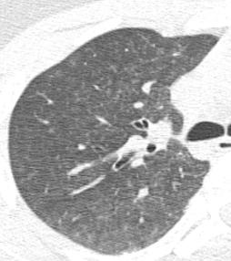
In the example above, the opacities are very small and difficult to see.

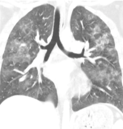
But they’re a little more obvious here!

Other CT findings include small pulmonary nodules in the upper lobes or along peripheral pulmonary vessels. These are thought to be areas of obstruction caused by the emboli. Nonspecific pleural effusions may be seen, and bronchial thickening has also been described. Rarely, fat globules may be seen in the lower extremity veins or IVC, and should immediately raise suspicion for developing FES even before symptoms develop.

CNS (60% of cases): If they occur, CNS changes generally crop up after the pulmonary manifestations begin. Generally, they start as mild confusion, but can progress to decreasing level of consciousness and even coma. Focal neurologic deficits are occasionally seen, and seizures can occur.

The actual mechanism behind this appears to be very similar to the skin changes which will be described in the next section. Emboli occur in vessels predominantly in the white matter of the brain. This leads to petechial hemorrhages, which are likely due to the inflammatory mechanisms previously described.

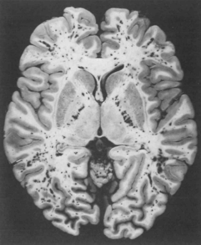
Note the numerous dark petechiae visible in the white matter in this specimen.

Retinal exam can also show evidence of fat embolism. Fat globules may actually be seen in the retinal vessels early.

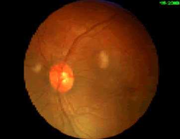
Note the fat globules at the 9:30 and 2:00 positions to the optic nerve in the image above.

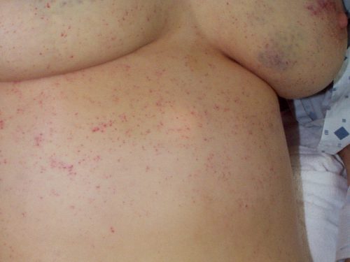
Skin (33% of cases): The most recognizable sign of FES is the petechial skin rash. This rash usually involves the torso, and axillary petechiae are very common. It can spread to involve the head and neck, and occasionally the extremities. Subconjunctival hemorrhages are sometimes seen. The rash tends to be transient and usually lasts only a few days. Here is an example of the classic petechial rash.

Other findings: Fat globules may be found in the urine in patients with FES. However, they are commonly present in patients with long bone fractures, so their presence is not helpful or predictive. Nonspecific findings such as fever, leukocytosis, anemia, and thrombocytosis are also relatively common. In severe cases, cardiac dysfunction, hypotension, and peripheral hypoperfusion can occur. I have personally seen necrosis of fingers and toes from a very severe case.

Unfortunately, the “classic” triad of mental status changes, skin rash, and pulmonary insufficiency are seen in only a small minority of patients. Typically, only one or two signs and symptoms appear at the same time, making diagnosis a bit challenging.

Tomorrow, making the diagnosis of fat embolism syndrome.

Fat Embolism vs Fat Embolism Syndrome

It’s fat embolism week! Fat embolism syndrome (FES) is one of those clinical problems that trauma professionals read about during their training, then rarely ever see. Although the clinical manifestations are frequently mild, they can progress rapidly and become life-threatening. Over the next five days, I’ll try to  help you better understand this condition, and provide details on diagnosis and treatment.

Fat embolism syndrome (FES) is a constellation of findings that arise from a single, unified cause: the escape of fat globules into the circulation (fat embolism). The ultimate resting places of those globules determine the specific manifestations of FES seen in clinical practice. When it occurs, it typically becomes apparent 24 to 72 hours after injury.

Simple fat embolism occurs to some degree any time tissues containing fat are manipulated or injured. It has been demonstrated during plastic surgical injections for cosmetic purposes and lipid infusions. It is more frequently seen with orthopedic injuries, especially those involving the femurs and pelvis. And it makes sense that the more fractures that are present, the more likely fat embolism will occur. Embolism is also known to occur when performing orthopedic procedures, particularly those involving the marrow cavity (intramedullary nailing), but has also been reported in total knee and hip procedures.

Fat embolism syndrome has a generally reported incidence of 1 – 10%, although I believe that is on the high side. I see a case every 3 – 4 years in a predominantly blunt, fracture-laden practice. Fat embolism without symptoms occurs much more frequently. A study from 1995 using transesophageal echo found evidence of emboli in 90% of patients with long bone fractures.

But how do these fat globules get into the circulation and produce such chaos? We know that they can be mechanically pushed into small venules when tissues containing fat cells or bone marrow are injured. In bone, there are numerous small venules located throughout that are anchored to it. When the bone is fractured, these venules tear and are held open so yellow (fatty) marrow can be pushed into them.

If enough emboli enter the blood stream, they may accumulate in the end vessels of tissues and block flow. Although this is a simple and appealing explanation, it may not be the full story. If the emboli primarily occur during and after injury, why does it take several days for the full-blown syndrome to develop?

A likely explanation is that the fat globules begin to degrade while in the circulatory system. Breakdown into free fatty acids results in the release of a cascade of cytokines and other mediators. The inflammatory response around the end vessels create the gross pathology that we associate with fat embolism syndrome.

Tomorrow, clinical manifestations of fat embolism syndrome.

Gluteal Compartment Syndrome

Compartment syndromes can occur virtually anywhere a muscle group is surrounded by relatively unforgiving soft tissue. In trauma, these classically involve the calf, forearm, and occasionally the thigh compartments. But they are occur unsuspected in the less common areas they can easily be missed, leading to significant morbidity, disability, and even death.

The gluteal compartment syndrome is one of those uncommon occurrences. Actually, it’s extremely rare, with less than 50 cases documented in the English literature. It is typically seen in patients who are impaired in some manner (drugs, alcohol, stroke) and are unable to move. If they lie in such a way that significant pressure is exerted on the buttock, the full syndrome can develop.

Typical symptoms include swelling, firmness, and pain in the buttock. Neurologic findings are fairly common. Paresthesias can develop late, and pressure on the sciatic nerve can ultimately begin to cause a sciatic palsy.

As with most compartment syndromes, the diagnosis is usually made solely on physical exam. However, in patients with more body fat it may not be as apparent. A pressure monitor can be inserted directly into the fleshiest part of the buttock, and elevated pressures (approaching or exceeding 30 torr) clinches the diagnosis.

The mainstays of treatment are surgical release and physiologic support, primarily for rhabdomyolysis and secondary renal injury. There are two types of incision that may be used. The classic straight line, shown on the right below, is simple but significantly disfiguring. The question mark incision on the left is kinder and gentler, but more challenging to perform properly.

Bottom line: Compartment syndromes can occur in any enclosed muscle group, which is just about all of them. Always be suspicious if your patient has unexplained elevations of CK, especially if they have tight muscle groups or deep pain in hard to access muscles. Err on the side of checking pressures and releasing those compartments in order to minimize morbidity and ultimate disability.

Reference: Gluteal compartment syndrome: a case report. Cases J. 2:190, 2009.

Air Embolism From an Intraosseous (IO) Line

IO lines are a godsend when we are faced with a patient who desperately needs access but has no veins. The tibia is generally easy to locate and the landmarks for insertion are straightforward. They are so easy to insert and use, we sometimes “set it and forget it”, in the words of infomercial guru Ron Popeil.

But complications are possible. The most common is an insertion “miss”, where the fluid then infuses into the knee joint or soft tissues of the leg. Problems can also arise when the tibia is fractured, leading to leakage into the soft tissues. Infection is extremely rare.

This photo shows the inferior vena cava of a patient with bilateral IO line insertions (black bubble at the top of the round IVC).

During transport, one line was inadvertently disconnected and probably entrained some air. There was no adverse clinical effect, but if the problem is not recognized and the line is not closed properly, there could be.

Bottom line: Treat an IO line as carefully as you would a regular IV. You can give anything through it that can be given via a regular IV: crystalloid, blood, drugs. And even air, so be careful!

Bullet In The Disk Space: Big Deal Or Not?

In an earlier post, I reviewed the problems with lead poisoning that can occur if a bullet remains in contact with a joint space / synovial fluid, or ends up in the GI tract. But what about if it comes to rest in an intervertebral joint space? They’re dry, right?

The first case report I could find dates back to 1981. A male presented to Parkland Memorial Hospital 12 years after a gunshot to the abdomen in which the bullet lodged in a disk space. He was treated for a GI bleed, but was also noted to have many signs and symptoms of high lead levels. These included irritability, anemia, headache, lethargy, muscle weakness and confusion. A blue line was noted on the gums. X-ray of the lumbar spine showed the bullet fragment in the center of the disk space, and a cystic mass in the prevertebral area that appeared radiodense as well. Blood lead levels were elevated. The patient underwent diskectomy, resection of the mass, chelation therapy, and recovered.

Another case report from 2010 was similar in many ways. The patient was young, had a gunshot 5 years previously, and presented with symptoms of lead poisoning. The appearance of the bullet in the disk space was similar to the last case, in that the bullet could be seen within it, and there appeared to be additional radiopaque material surrounding it. It almost looked like lead was flowing out of the bullet into the disk. This case was also treated with surgical removal and chelation with a successful result.

A literature review was conducted 15 years ago that examined other case reports of bullets in the spine. Over a 25-year period 238 patients were identified with this injury. Only 12 had bullets or fragments in the disk space. All were tested for plumbism, and only one was positive. He underwent diskectomy and resection with resolution of the high lead levels.

Bottom line: We know that a bullet in contact with synovial fluid is bad, with rapid leaching of lead into the circulation. There are also suggestions that lead in contact with CSF can cause a similar problem. However, the intervertebral disk space is usually considered to be “dry” and doesn’t usually cause a problem.

However, patients with a bullet in this location should be cautioned that they do have a small risk of developing lead poisoning. They should be tested about six months post-injury to see if lead levels are on the rise. They should also be cautioned to report the development of new back pain. Structural disruption by the bullet may slowly lead to anatomic changes that result in chronic pain. And be very suspicious if there is radiopaque material in the disk space in addition to the bullet itself!

References:

  1. Acute lead intoxication from a bullet in the intervertebral disk space. JBJS 63A(7):1180-1182, 1981.
  2. Lead Poisoning by Intradiscal Firearm Bullet. Spine 35(4):E140-E143, 2010.
  3. Long-Term Clinical Manifestations of Retained Bullet
    Fragments Within the Intervertebral Disk Space. J Spinal Disord Tech 17(2):108-111, 2004.