Determining The Age Of Bruises

On occasion, you may encounter a patient who has bruising and wonder how old the injuries are. Or there may be several bruises and you would like to know if they occurred at different times. This becomes especially important when dealing with injured children in whom there is a suspicion of abuse.

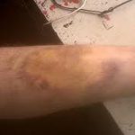
Bruising occurs when blood leaks from blood vessels into the skin and subcutaneous tissues. If the skin and soft tissues are firm, bruising is not as apparent. In areas where the skin and soft tissues are loose, such as the peri-orbital areas and scrotum, bruising is visible early and may be extensive. The elderly tend to bruise easily because both the skin and subcutaneous tissue are very thin and friable.

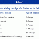
A predictable series of color changes occurs with most bruises. During the acute phase, the color is usually reddish and the area may be raised and tender. After about 2 days, the color turns purple and any swelling usually disappears. Over the next week, the color changes to green and yellow as the heme metabolizes. Finally, the color fades and by two weeks most evidence of the injury is gone.

The table above is a key to estimating bruise age. However, this is not an exact science! A number of studies have been performed showing that examiners given photographs of bruises of various ages were not terribly accurate. Fresh and intermediate bruises were identified fairly accurately, but not so for older bruises.

The trauma professional may find it helpful to use these guidelines when trying to decide if there are both fresh and older bruises present. This may indicate that an older adult may be suffering from frequent falls, or that a child needs to be evaluated for abuse.

Reference: Estimation of the age of bruising. Arch Dis Childhood 74:53-55, 1996.

Bucket Handle Injury – Part 2

Yesterday, I wrote about the basics of bucket handle injuries of the intestine. Today, I’ll deal with diagnosing them.

An understanding of the mechanism of injury and a good physical exam are paramount. If the patient took a significant blow to the abdomen, especially in a motor vehicle crash (lap belt), be very suspicious. Any abdominal pain is of concern, particularly in the right lower quadrant (most common injury location). If a CT is indicated and there are focal changes in the mesenery or bowel wall, a trip to the OR is advised.

In some patients, the bowel is devascularized and takes 2-3 days to become necrotic. They experience slowly increasing focal pain, and once this develops it’s time to go to the operating room.

Intubated and/or comatose patients can be problematic in making this diagnosis. There is no physical exam, so the trauma professional has to rely on surrogates. The white blood cell (WBC) count is very helpful. The WBC count is typically elevated into the 15,000-20,000 range immediately after trauma, and declines to normal within about 12 hours. If it begins to climb again after 24 hours, especially if it exceeds 20,000, an intestinal injury is likely.

CT scan and abdominal ultrasound are also helpful. A repeat CT scan may show a change in the volume of fluid, or a change in its character. If the amount of fluid increases significantly, or if a fluid bi-layer is seen, a bucket handle injury is very likely. These findings are pertinent in awake patients as well, but the physical exam usually makes use of these diagnostics unnecessary.

Related posts:

Bucket Handle Injury – Part 1

Bucket Handle Injury

A bucket handle injury is a type of mesenteric injury of the intestine. The intestine itself separates from the mesentery, leaving a devascularized segment of bowel that looks like the handle on a bucket (get it?).

These injuries can occur after blunt trauma to the abdomen. The force required is rather extreme, so the usual mechanism is motor vehicle crash. In theory, it could occur after a fall from a significant height, and I have seen once case where a wood fragment was hurled at the abdomen by a malfunctioning lathe.

The mechanics of this injury are related to fixed vs mobile structures in the abdomen. Injuries tend to occur adjacent to areas of the intestine that are fixed, such as the cecum, ligament of Treitz, colonic flexures and rectum. During sudden deceleration, portions of the intestine adjacent to these areas continue to move, pulling on the nearby attachments. This causes the intestine itself to pull off of its mesentery.

The terminal ileum is the most common site for bucket handle tears. Proximal jejunum, transverse colon, and sigmoid colon are other possible areas. The picture above shows multiple bucket handle injuries in one patient. There are 3 injuries in the small bowel, and one involving the entire transverse colon. Note the obviously devascularized segment at the bottom center of the photo.

Always think about the possibility of this injury in patients with very high speed decelerations. Seat belt marks have a particularly high association with this injury. If your patient has an abnormal exam in the right lower quadrant, or if the CT shows unusual changes there (“dirty” mesenteric fat, thickened bowel wall, extravasation), I recommend a trip to the OR. In these cases, an injury will nearly always be present.

Tomorrow: These injuries can be subtle in an awake patient with a reliable exam. On Friday I’ll write about how you can detect it in unconscious patients.

Source: personal archive. Not treated at Regions Hospital

There will not be a scheduled post today or tomorrow. We are undergoing an American College of Surgeons reverification visit for our adult and pediatric Level I trauma centers today. Things are a bit busy, but I’ll resume on Thursday!

Yes, Smoking is Bad!

Everybody knows that smoking is bad. But how often have you stopped by to see one of your trauma patients and have been told “they’re out smoking?” Well, it turns out it’s bad for their injuries as well.

A German group looked at the effects of smoking on healing of a “simple” tibial fracture. They looked at 103 patients who underwent treatment for an isolated tibial shaft fracture at a trauma center. Patients with more complicated problems like extension into a joint, open fracture (Gustilo III), or significant soft tissue injury were excluded. 

Patients were divided into non-smokers and smokers (including previous smokers). A total of 85 patients were studied, and there were roughly half in each group. The nonsmoking group experienced no delayed or non-unions of their fractures. The smoking group reported 9 delayed unions and 9 non-unions in 46 patients! As expected, time off work and eventual functional outcome was worse as well.

Bottom line: The exact mechanism for impairment of fracture healing by smoking is unclear. It may be due to physiologic effects of inhaled tobacco components on blood flow, blood vessels, transforming growth factor levels or collagen formation. It could also be a secondary effect of socioeconomic variables, patient compliance, or a host of other factors. Regardless, it’s bad. Smoking should be forbidden while in hospital, and should be strongly discouraged after discharge.

Reference: Cigarette smoking influences the clinical and occupational outcome of patients with tibial shaft fractures. Injury 42:1435-1442, 2011.

Home of the Trauma Professional's Blog

Do you want to get a daily email every time there’s a new post? See what I’m up to.

Click here to get details and subscribe!

[accua-form fid=”1″]

[mc4wp_form id=”2023″]