Pop Quiz: The Answer!

There were lots of interesting guesses regarding this photo! Some were very creative, and thought I might be throwing a curve ball. Alas, this was much more straightforward.

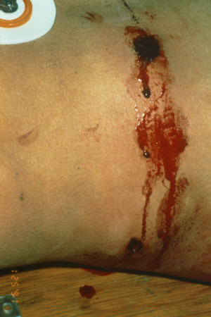
What you see is a pair of wounds located just at or slightly above the iliac crest on the right side. If you look carefully, you will see a powder burn around the anterior wound, indicating a close range gunshot. So this would appear to be a run of the mill gunshot to the abdomen; just run to the OR, right?

Not so fast! There are some nuances when dealing with this type of wound. The first things to look at are the vital signs. If they’re not stable, then there is major bleeding present and the patient needs to go to the OR now. Next, do a good exam. As always, stick to the ATLS protocol to make sure you’re not focusing on the abdomen and missing other significant findings. If the abdominal exam is abnormal (tenderness, peritoneal signs) there is either bleeding or contamination and once again it’s time to go to OR. About 98 times out of 100, that’s where you’ll be with a picture like this.

However, if you’ve gotten to this point with none of the above, there is the small possibility that this might be a tangential injury. The flanks (“love handles”) tend to be fairly fatty in some men, especially the obese. And since most civilian gunshots are low velocity, there is less likelihood of deeper injury from blast effect. Local wound exploration is tough in this area due to the amount of fat and the deeper musculature. 

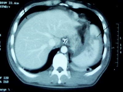
My preferred method for evaluating this (rare) type of patient is a quick CT scan of the abdomen and pelvis. The pelvic part is important, because you are looking for obvious penetration and blood in the pelvis. If you see either, it’s time to head to the OR. Very rarely (on the right side) you may see a contusion or superficial laceration of the liver, meaning that there was penetration. However, if there is no possible way the bowel was injured, it is acceptable to closely observe the patient.

Oh, and the board? Back in the day before everything was made of plastic, they actually made backboards out of fairly nice wood!

Pop Quiz!

Okay, here’s a picture of an injury. First, what happened? Then, what’s your management approach? And why are they lying on a piece of wood?!

It’s not as straightforward as you might think! Answer tomorrow.

Source: personal archive. Not treated at Regions Hospital.

Undergrading Spleen Injury

We love our CT scans! They’re so high tech, with such detailed images popping up on the monitor so quickly. To take advantage of the detail, we’ve come up with fancy grading systems that can be used to direct care. But are they all they’re cracked up to be?

CT grading of spleen injury is a prime example. We’ve got a nice, detailed system that looks at laceration depth, subcapsular hematoma size and vascular injury. We can use it to predict the likelihood of needing an operation and where we should admit someone in the hospital (ICU vs ward). And when we see the injury on the screen, we believe that we can accurately apply the scoring system to these beautiful images.

But unfortunately, it’s not that simple.Scanning obtains multiple images in an axial plane and lays them out for us to look at. However, the spleen (and most other organs) and not shaped like a cube. It is curved, with complex nooks and crannies that can look like cracks. Moderate to large hematoma around the spleen can obscure lacerations. And the hilum is even more complicated and variable in shape.

Because of this, CT scans of the abdomen tend to underestimate the true extent of injury, especially in the higher grades. Grade I and II injuries are usually accurate, but in Grades III-V, the scan tends to undergrade by 1 (30% of cases) or 2 grades (45% of cases) when re-graded at surgery.

Bottom line: Grade I and II injuries are generally managed in a lower intensity setting and almost never require operation. But beware of the higher grades! It is very likely that it’s higher than you think. This means that if your patient slowly becomes tachycardic or their blood pressure softens, believe the clinical evidence. Don’t rely on a CT scan that was done hours ago that may be hiding a more severe injury than you think! (This applies to liver injuries as well)

Related posts:

Reference: Correlation of operative and pathological injury grade with computed tomographic grade in the failed nonoperative management of blunt splenic trauma. Euro J Trauma Emerg Surg – Online First 2 Mar 2012.

What Percent Pneumothorax Is It?

What percent pneumothorax?

Frequently, radiologists and trauma professionals are coerced into describing the size of a pneumothorax seen on chest xray in percentage terms. They may say something like “the patient has a 30% pneumothorax.”

The truth is that one cannot estimate a 3D volume based on a 2D study like a conventional chest xray. Everyone has seen the patient who has no or a minimal pneumothorax on a supine chest xray, only to discover one of significant size with CT scan.

Very few centers have or use the software that can determine the percentage of chest volume taken up with air. There are only two percentages that can be determined by viewing a regular chest xray: 0% and 100%. Obviously, 0% means no visible pneumothorax, and 100% means complete collapse. Even 100% doesn’t really look like 100% because the completely collapsed lung takes up some space. See the xray at the top for a 100% pneumothorax.

If you line up 10 trauma professionals and show them a chest xray with a pneumothorax, you will get 10 different estimates of their size. And there aren’t any guidelines as to what size demands chest tube insertion and what size can be watched.

Bottom line: The solution is to be as quantitative as possible. Describe the pneumothorax in terms of the maximum distance the edge of the lung is from the inside of the chest wall, and which intercostal space the pneumothorax extends to. So instead of saying “the patient has a 25% pneumo,” say “the pneumothorax is 1 cm wide and extends from the apex to the fifth intercostal space on an upright film.”

Home of the Trauma Professional's Blog

Do you want to get a daily email every time there’s a new post? See what I’m up to.

Click here to get details and subscribe!

[accua-form fid=”1″]

[mc4wp_form id=”2023″]