The Pan-Scan For Trauma

Diagnostic imaging is a mainstay in diagnosing injuries in major trauma patients. But the big questions are, how much is enough and how much is too much? X-radiation is invisible but not inocuous. Trauma professionals tend to pay little attention to radiation that they can’t see in order to diagnose things they can’t otherwise see. And which may not even be there.

There are two major camps working in emergency departments: scan selectively and scan everything. It all boils down to a balance between irradiating enough to be satisfied that nothing has been missed, and irradiating too much and causing harm later. 

A very enlightening study was published last year from the group at the University of New South Wales. They prospectively looked at their experience while moving from selective scanning to pan-scanning.They studied over 600 patients in each cohort, looking at radiation exposure, missed injuries, and patient injury and discharge disposition variables.

Here are the interesting findings:

  • Absolute risk of receiving a higher radiation dose increased from 12% to 20%. This translates to 1 extra person of every 13 evaluated receiving a higher dose.
  • The incidence of receiving >20 mSv radiation dose nearly doubled after pan-scanning. This is the threshold at which we believe that cancer risk changes from low (<1:1000) to moderate (>1:1000).
  • The risk of receiving >20 mSv was lower in less severely injured patients (sigh of relief)
  • There were 6 missed injuries with selective scanning and 4 with pan-scanning (not significant). All were relatively minor.

Bottom line: Granted, the study groups are relatively small, and the science behind radiation risk is not very exact. But this study is very provocative because it shows that radiation dose increases significantly when pan-scan is used, but there was no benefit in terms of decreased missed injury. If we look at the likelihood of being helped vs harmed, patients are 26 times more likely to be harmed in the long term as they are to be helped in the short term. The defensive medicine naysayers will always argue about “that one catastrophic case” that will be missed, but I’m concerned that we’re creating some problems for our patients in the distant future that we are not worrying enough about right now.

Related posts:

Reference: Comparison of radiation exposure of trauma patients from diagnostic radiology procedures before and after the introduction of a panscan protocol. Emerg Med Australasia 24(1):43-51, 2012.

If A Tree Falls In A Forest…

Time for a little philosophy today. There seem to be two camps in the world of initial diagnostic testing for trauma: selective scanning vs scan everything. I admit that I am one of the former. Yes, the more tests you do, the more things you will find. Some will be red herrings. Some may be true positives, but are they important? Here’s the key question:

“If a tree falls in a forest and no one is around, does it make a sound?”

There is a clinical corollary to this question in the field of trauma:

“If an injury exists but no one diagnoses it, does it make a difference (if there would be no change in treatment)?”

Here’s an example. On occasion, my colleagues want to order diagnostic studies that won’t make any clinical difference, in my opinion. A prime example is getting a chest CT after a simple blunt assault. A plain chest xray is routine, and if injuries are seen or the physical exam points to certain diagnoses, appropriate interventions should be taken. But adding a chest CT does not help. Nothing more than the usual pain management, pulmonary toilet, and an occasional chest tube will be needed, and those can be determined without the CT.

Trauma professionals need to realize that we don’t need to know absolutely every diagnosis that a patient has. Ones that need no treatment are of academic interest only, and can lead to accidental injury if we look for them too hard (radiation exposure, contrast reaction, extravasation into soft tissues to name a few). This is how we get started on the path to “defensive medicine.”

Bottom line: Think hard about every test you order. Consider what you are looking for, what you might find, and if it will change your management in any way. If it could, go ahead. But always consider the benefits versus the potential risks, or what I call the “juice to squeeze ratio.”

Tomorrow I’ll look at some of the “scan all” vs “scan selectively” literature. Which camp are you in?

References:

  • George Berkeley, A Treatise Concerning the Principles of Human Knowledge, 1734, section 45.
  • paraphrased by William Fossett, Natural States, 1754.

Do You Really Need To Repeat That Xray?

It happens all the time. You get that initial chest and/or pelvic xray in the resuscitation room while evaluating a blunt trauma patient. A few minutes later the tech returns with another armful of xray plates to repeat them. Why? The patient was not centered properly and part of the image is clipped.

Do you really need to go through the process of setting up again, moving the xray unit in, watching people run out of the room (if they are not wearing lead, and see my post below about how much radiation they are really exposed to), and shooting another image? The answer to the question lies in what you are looking for. Let’s address the two most common (and really the only necessary) images needed during early resuscitation of blunt trauma.

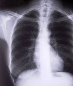
First, the chest xray. You are really looking for 3 things:

  • Big air (pneumothorax)
  • Big blood (hemothorax)
  • Big mediastinum (hinting at aortic injury)

Look at the clipped xray above. A portion of the left chest wall is off the image. If there were a large pneumothorax on the left, would you be able to see it? What about a large hemothorax? And the mediastinum is fully included, so no problem there. So in this case, no need to repeat immediately.

The same thing goes for the pelvis. You are looking for gross disruption of the pelvic ring, especially posteriorly because this will cause you to intervene in the ED (order blood, consider wrapping the pelvis). So if parts of the edges or top and bottom are clipped, no big deal.

Bottom line: Don’t let the xray tech disrupt the team again by reflexively repeating images that are not technically perfect. See if you can use what you already have.  And how do you decide if you need to repeat it later, if at all? Consider the mechanism of injury and the physical exam. Then ask yourself if there is anything you could possibly see that was not imaged the first time that would change your management in any way. If not, you don’t need it. But it certainly will irritate the radiologists!

Related posts: 

Pop Quiz Answer – Jet Ski Injury

This pop quiz involved a young woman who fell from a jet ski at high speed and was initially okay. Later that day, she developed lower abdominal pain and sought evaluation in your ED. There were a number of thoughtful answers via the comments section and Twitter.

According to the First Law of Trauma, the pain is related to the mishap until proven otherwise. You must approach it like any moderate speed motor vehicle crash. In many ways, this mechanism is similar to a motorcycle or bicycle crash, without the road rash. However, high speed and water can also combine to cause a unique injury, the so called water ski / jet ski douche and enema. This occurs when the rider enters the water with a significant feet first component, causing a jet of water to be forced into the vagina or rectum.

As always though, start with a thorough history and physical exam. In this case, the patient has diffuse lower abdominal tenderness, but no other findings on exam. Because of the possibility of water jet injury, a thorough exam of both vagina and rectum is indicated. This requires a speculum and anoscope. Any anomalies that are noted are an indication to proceed to the OR for a thorough exam under anesthesia.

Blunt abdominal injury is also a concern, so FAST may be performed. However, the abdominal pain is an indication for abdominal/pelvic CT using our blunt trauma imaging protocol. A solid organ injury can be managed in the usual manner. But if any anomalies other than the trace pelvic fluid occasionally seen in young women is present, the patient must go to OR.

If the patient does need an operation, start with vaginal and rectal exams again, under anesthesia. Most vaginal lacerations are small and easily closed. However, there have been reports of extensive laceration with heavy bleeding. Simple rectal tears may be repaired, but more complicated ones may also require fecal diversion. If the injury appears complex, a laparotomy will be necessary, and diversion with a colostomy will usually be required.

Bottom line: This injury is an example of what I call a two-factor mechanism: blunt trauma plus high pressure injection in this case. The trauma professional needs to recognize both and resist the temptation to focus on the more obvious one. Think through the evaluation and management algorithms for each one, combining them where appropriate.

Related posts:

Home of the Trauma Professional's Blog

Do you want to get a daily email every time there’s a new post? See what I’m up to.

Click here to get details and subscribe!

[accua-form fid=”1″]

[mc4wp_form id=”2023″]