The “Passing” Of The Rectal Exam In Trauma

It has long been standard operating procedure to perform a digital rectal exam in all major trauma patients. The belief has always been that valuable information about blood in the GI tract, the status of the urethra, and the neuro exam (rectal tone) could be gleaned from the exam.

Unfortunately, the exam also serves to antagonize or even further traumatize some patients, especially those who may be intoxicated to some degree. On a number of occasions I have seen calm patients become so agitated by the rectal that they required intubation for control.

So is it really necessary? A study in 2001 conducted over a 6 month period (1) showed that the rectal exam influenced management in only 1.2% of cases. The authors felt that there was some utility in 3 special cases:

  • Spinal cord injury – looking for sacral sparing
  • Pelvic fracture – looking for bone shards protruding into the rectum
  • Penetrating abdominal trauma – looking for gross blood

A more recent 2005 study (2) was also critical of the rectal exam and found that using “other clinical indicators” (physical exam and other diagnostic study information) was at least equivalent, changing management only 4% of the time. They concurred with the first two indications above as well.

The Bottom Line: For most major trauma patients, the rectal exam is not worth the patient aggravation it causes. I still recommend it for the 3 special cases listed above, however, as there are no equivalent exams for these potentially serious patient problems. And remember, DON’T do it while the patient is in the logroll position. No patient likes a rectal exam, so they’ll do their best to defeat your attempt at spine precautions if you have them on their side. Supine, frog-leg only.

References:
1. Porter, Urcic. Am Surg. 2001 May;67(5):438-41.
2. Esposito et al. J Trauma. 2005 Dec;59(6):1314-9.

Stuff You Sterilize Other Stuff With May Not Be Sterile??

When one works in the trauma field, or medicine in general, we deal with the need for sterility all the time. We use equipment and devices that are sterile, and we administer drugs and fluids that are sterile. In surgery, we create sterile fields in which to use this sterile stuff.

In the past few years, we’ve come to the realization that the sterility we take for granted may not always be the case. There have been several cases of contaminated implanted hardware. And most recently, supposedly sterile injectable steroids were found to be contaminated with fungus, leading to several fatal cases of meningitis.

A recent article in the New England Journal of Medicine brings a bizarre problem to light: microbial stowaways in the topical products we use to sterilize things. Most drugs and infused fluids are prepared under sterile conditions. However, due to the antimicrobial activity of topical antiseptics, there is no requirement in the US that they be prepared in this way.

A number of cases of contamination have been reported over the years:

  • Iodophor – contamination with Burkholderia and Pseudomonas occurred during manufacture, leading to dialysis catheter infection and peritonitis
  • Chlorhexidine – contaminated with Serratia, Burkholderia and Ralstonia by end users, leading to wound infections, catheter infections, and death
  • Benzalkonium chloride – contaminated with Burkholderia and Mycobacteria by end users, causing septic arthritis and injection site infections

Bottom line: Nothing is sacred! This problem is scarier than you think, because our most basic assumptions about these products makes it nearly impossible for us to consider them when tracking down infection sources. Furthermore, they are so uncommon that they frequently may go undetected. The one telltale sign is the presence of infection from weird bacteria. If you encounter these bugs, consider this uncommon cause. Regulatory agencies need to get on this and mandate better manufacturing practices for topical antiseptics.

Reference: Microbial stowaways in topical antiseptic products. NEJM 367:2170-2173, Dec 6 2012.

Trauma Mythbusters: NSAIDs And Fracture Healing

Trauma hurts like hell. Over the years, we’ve developed quite a few ways of combating this pain. A number of drug classes have been developed to reduce it. One of the more common non-narcotic drug classes are the NSAIDs. As I’ve mentioned before, every drug has dozens of effects. Drug companies market a particular medication based on one of the predominant effects. All the others are considered side effects.

NSAIDs are not unique; they have lots of side effects as well. In 2003, several papers brought to light possible interactions between these drugs and fracture healing. Specifically, there were questions about these drugs interfering with the healing process and of increasing the number of delayed unions or nonunions. But once again, how convincing were these papers, really?

It would seem to make sense that NSAIDs could interfere with bone healing. This  process relies heavily on the regulation of osteoblast and osteoclast function, which itself is regulated by prostaglandins. Since prostaglandins are synthesized by the COX enzymes, COX inhibitors like the NSAIDs should have the potential to impair this process. Indeed, animal studies in rats and rabbits seem to bear this out.

But as we have seen before, good animal studies don’t always translate well to human experience. Although a study from 2005 suggested that NSAID administration in older patients within 90 days of injury had a higher incidence of fracture nonunion, the study design was not a very good one. It is equally likely that patients who required these drugs in this age group may have been at higher risk for nonunion in the first place.

In fact, there are no large, prospective randomized studies that have explored the effect of short-term or long-term NSAID administration on fracture repair. But there have been several smaller studies that showed absolutely no effect on nonunion with short-term administration of this drug class. Yet the dogma that leads us to avoid giving these drugs persists.

Bottom line: Once again, the animal data is clear but the human data is not. Although there are theoretical concerns about their use, there is not enough solid risk:benefit information to abandon short-term NSAID use in patients who really need them. NSAIDs can and should be prescribed in patients with short-term needs and simple fractures.

References:

  1. Effects of nonsteroidal anti-inflammatory drugs on bone formation and soft-tissue healing. J AM Acad Orthop Surg 12:139-43, 2004.
  2. Effect of COX-2 on fracture-healing in the rat femur. J Bone Joint Surg Am 86:116-123, 2004.
  3. Effects of perioperative anti-inflammatory and immunomodulating therapy on surgical wound healing. Pharmacotherapy 25:1566-1591, 2005.
  4. Pharmacological agents and impairment of fracture healing: what is the evidence? Injury 39:384-394, 2008.
  5. High dose nonsteroidal anti-inflammatory drugs compromise spinal fusion. Can J Anaesth 52:506-512, 2005.

Handlebar Injury Case: The Answer

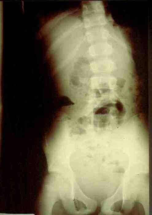
Okay, time for the answer. This 12 year old crashed his moped, taking handlebar to the mid-epigastrium. Over the next 3 days, he felt progressively worse and finally couldn’t keep food down.

Mom brought him to the ED. The child appeared ill, and had a WBC count of 18,000. The abdomen was firm, with involuntary guarding throughout and a hint of peritonitis. The diagnosis was made on the single abdominal xray shown yesterday. A closeup of the good stuff is above.

Emergency docs, your differential diagnosis list with this history is a pancreatic vs a duodenal injury based on the mechanism.

Classic findings for duodenal injury:

  • Scoliosis with the concavity to the right. This is caused by psoas muscle irritation and spasm from retroperitoneal soiling by the duodenal leak.
  • Loss of the psoas shadow on the right. Hard to see on this xray, but the left psoas shadow is visible, the right is not. This is due to fluid and inflammation along this plane.
  • Air in the retroperitoneum. In this closeup, you can actually see tiny bubbles of leaked air outlining the right kidney. There are also bubbles along the duodenum and a few along the right psoas.

We fluid resuscitated first (important! dehydration is common and can lead to hemodynamic issues upon induction of anesthesia) and performed a laparotomy. There was a  blowout in the classic position, at the junction of 1st and 2nd portions of the duodenum. The hole was repaired in layers and a pyloric exclusion was performed, with 2 closed drains placed in the area of the leak.

The child did well, and went home after 5 days with the drains out. Feel free to comment or leave questions!

Traveling In France!

I’ll be travelling through France for the next two weeks, stopping by a few hospitals to visit, I hope. France is in my top 10 list of international readers. And interestingly, most of my French readers are not in Paris! 

Most of my posts will be “Best Of” while I am away. Additionally, there will not be a Trauma MedEd newsletter this month. But I’ll make it up to you in June!

Follow my progress on FourSquare and Twitter! I hope to meet some of my international readers out there! Tweet me if I’m in your neighborhood.

Michael

Home of the Trauma Professional's Blog

Do you want to get a daily email every time there’s a new post? See what I’m up to.

Click here to get details and subscribe!

[accua-form fid=”1″]

[mc4wp_form id=”2023″]