All posts by The Trauma Pro

Where Are All The Pediatric Trauma Centers?

I have constructed this map from available resources from the American College of Surgeons and numerous state agencies. ACS verified pediatric centers have a diamond in their icon; Level I is red and Level II is yellow. Non-ACS centers are pink (Level I) or blue (Level II). The Level I pediatric center at Regions Hospital is the green star.

I have made every attempt at accuracy, but things do change. If I have omitted any centers or misclassified them, please leave a comment or email me!

Inline Stabilization vs Inline Traction of the Cervical Spine

Members of the trauma team must frequently protect the cervical spine when moving the patient or performing certain procedures. In most cases, a cervical collar is placed which does a fine job of this. Occasionally, though, the collar must be removed to provide access to areas near or under the collar.

When the collar is off, someone must be charged with immobilizing the cervical spine. Sometimes this is incorrectly referred to as providing inline traction and not inline stabilization.There is a big difference!

Inline traction is used to try to realign cervical vertebra that are malpositioned due to fracture or ligamentous injury. This should only be performed under the guidance of a neurosurgeon!

Inline stabilization merely means that the patient (or trauma professional) is restrained from moving the cervical spine. This is commonly needed while intubating the patient, so that the intubator does not extend the neck when trying to visualize the cords.

Why is this important? Check out the images below. If a severe injury has already occurred, traction on the neck may have devastating consequences! Inline stabilization is the only way to go.

Spine injury AO dissociation

How Much Radiation is the Trauma Team Really Exposed To?

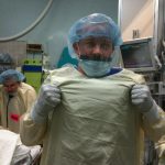
Okay, so you’ve seen “other people” wearing perfectly good lead aprons lifting them up to their chin during portable xrays in the trauma bay. Is that really necessary, or is it just an urban legend?

After hitting the medical radiation physics books (really light reading, I must say), I’ve finally got an answer. Let’s say that the xray is taken in the “usual fashion”:

  • Tube is approximately 5 feet above the xray plate
  • Typical chest settings of 85kVp, 2mAs, 3mm Al filtration
  • Xray plate is 35x43cm

The calculated exposure to the patient is 52 microGrays. Most of the radiation goes through the patient onto the plate. A very small amount reflects off their bones and the table itself. This is the scatter we worry about.

So let’s assume that the closest person to the patient is 3 feet away. Remember that radiation intensity diminishes as the square of the distance. So if the distance doubles, the intensity decreases to one fourth. By calculating the intensity of the small amount of scatter at 3 feet from the patient, we come up with a whopping 0.2 microGrays. Since most people are even further away, the dose is much, much less for them.

Let’s put it perspective now. The background radiation we are exposed to every day (from cosmic rays, brick buildings, etc) amounts to about 2400 microGrays per year. So 0.2 microGrays from chest xray scatter is less than the radiation we are exposed to naturally every hour!

The bottom line: unless you need to work out you shoulders and pecs, don’t bother to lift your lead apron every time the portable xray unit beeps. It’s a waste of time and effort!

Trauma 20 Years Ago: Early vs Delayed Femur Fixation

Today, we take for granted that fixing fractures early is a good thing. However, this topic was still under debate 20 years ago. Trauma care has always been prioritized, with life-threatening injuries taking precedence. It was very common for major trauma patients to undergo operation for their torso injuries, and then be deemed “too unstable” to undergo repair of their extremities.

Weigelt et al reported decreased pulmonary complications with early fixation in 1989. A study published in July 1990 looked at 121 early vs 218 late femur fixations with respect to more concrete outcomes. The patients were similar with respect to hypotension, transfusions and associated injuries.

They found that delayed fixation increased pulmonary shunt, especially in patients with more severe injuries, and increased the incidence of pneumonia in older patients. It also resulted in more ICU days and a significantly longer hospital stay in the more severely injured group.

This paper was a valuable addition that began to shape our appreciation for the importance of early fixation of most fractures. Major trauma makes patients sick, but they are in the best condition they will be in for weeks at the time they arrive at the hospital. This makes it the ideal time to take care of injuries that may otherwise contribute to morbidity and mortality.

Reference: Fabian et al. Improved outcome with femur fractures: early vs delayed fixation. J Trauma 30(7):792, 1990.